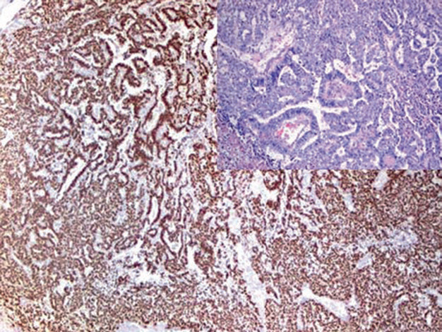
Figure 1. Strong nuclear expression of SALL-4 in yolk sac tumor.
In the interpretation of specific cases, it should be noted that SALL-4 is not only expressed in germ cell tumors but can also be expressed in some non-germ cell tumors, such as ovarian serous carcinoma, lung adenocarcinoma, cholangiocarcinoma, urothelial carcinoma, and small cell carcinoma.
Positive Expression Pattern: Nuclear
Recommended Positive Control Tissue: Seminoma
OCT-4 is expressed in early embryonic cells and plays an important role in the differentiation of pluripotent stem cells. High expression of OCT-4 is characteristic of seminoma and embryonal carcinoma, while spermatocytic seminoma does not express it. Additionally, in intratubular germ cell neoplasia, most atypical cells show nuclear expression of OCT-4, but non-neoplastic testicular cells do not. Therefore, this marker is helpful and specific for the diagnosis of intratubular germ cell neoplasia.

Figure 2. Positive nuclear expression of OCT-4 in tumor cells of seminoma.

Figure 3. Expression of OCT-4 in tumor cells of intratubular germ cell neoplasia.
It should be noted that some non-small cell lung cancers and breast cancers may express OCT-4; also, some cases of testicular and extratesticular diffuse large B-cell lymphoma may express this marker.
Positive Expression Pattern: Cell Membrane
Recommended Positive Control Tissue: Seminoma
PLAP is a membrane-associated glycoprotein mainly expressed in placental syncytiotrophoblasts from the 8th week of gestation. Some germ cell tumors can express this marker, such as seminoma, dysgerminoma, embryonal carcinoma, yolk sac tumor, and gonadoblastoma. Since PLAP is not specific to any particular germ cell tumor, when differentiating PLAP-positive germ cell tumors, a panel of markers should be used in combination.
Rarely, non-germ cell tumors may also show aberrant expression of PLAP, such as breast cancer and lung cancer. Additionally, it has been reported that tumors with myogenic differentiation may show cytoplasmic staining for PLAP, specifically such as embryonal rhabdomyosarcoma and smooth muscle tumors.
Positive Expression Pattern: Nuclear
Recommended Positive Control Tissue: Seminoma
SOX-2 is a transcription factor that forms a trimeric complex with OCT-4 at the DNA level and controls the expression of many genes during the embryonic development of the respiratory system, nervous system, and germ cells. For germ cell tumors, SOX-2 shows strong nuclear expression in embryonal carcinoma but is negative in seminoma, yolk sac tumor, and choriocarcinoma. SOX-2 can also be expressed in glial tumors of the brain and supratentorial PNET. In addition, SOX-2 expression can be seen in some lung squamous cell carcinomas and adenocarcinomas. There are also reports of SOX-2 positivity in some neuroendocrine carcinomas.
D2-40 is a type I transmembrane mucin, which will be discussed in detail in the subsequent articles of this series on vascular tumors. D2-40 is extremely helpful for the diagnosis of seminoma because it is negative in other germ cell tumors. Since D2-40 is positive in both tumor cells of seminoma and lymphatic vessels, it can also serve as a useful marker for lymphovascular invasion.
Positive Expression Pattern: Cytoplasmic
Recommended Positive Control Tissue: Placenta
HCG is a hormone produced by syncytiotrophoblasts, consisting of alpha and beta chains. The alpha chain shares the same amino acid sequence with other hormones such as LH, FSH, TSH, etc., while the beta chain has a unique structure and is more specific for syncytiotrophoblasts and related tumors. It should be noted that low-level expression of β-HCG can also occur in non-syncytiotrophoblastic tumors, such as lung cancer, colon cancer, and rarely lymphoma. Generally, expression of β-HCG in non-trophoblastic tumors indicates aggressive biological behavior. CD30 has been discussed in detail in the lymphoma section of this series. This marker is important in the diagnosis of Hodgkin lymphoma and anaplastic lymphoma. Additionally, CD30 positivity is characteristic of embryonal carcinoma. Rarely, weak positive staining for CD30 may occur in yolk sac tumors, which should be noted in the differential diagnosis of mixed germ cell tumors.

Figure 4. Strong positive expression of CD30 in embryonal carcinoma.
Positive Expression Pattern: Cytoplasmic
Recommended Positive Control Tissue: Granulosa Cell Tumor/Adrenal
Inhibin is a member of the transforming growth and differentiation factor family. It is a glycoprotein hormone composed of alpha and beta subunits, expressed in the adrenal glands and gonads. Its function is to inhibit FSH secretion and stimulate androgen synthesis in ovarian theca cells. Antibodies against inhibinα, anti-Müllerian hormone, and Melan A are important markers for the diagnosis of sex cord tumors. In ovarian surface epithelial-stromal tumors, seminoma, and embryonal carcinoma, inhibinαand anti-Müllerian hormone are negative. It should be noted that inhibinαand Melan A can also be expressed in other tumors, mainly adrenal cortical tumors.
Anti-Müllerian hormone belongs to the transforming growth factor-beta gene family. Its expression is regulated by SF-1, GATA, DAX1, and FSH. Anti-Müllerian hormone regulates male sexual differentiation by inhibiting Müllerian duct formation and preventing the transformation of Müllerian ducts into the uterus, fallopian tubes, and other Müllerian structures, and plays an important role in testicular differentiation. Without anti-Müllerian hormone, the Müllerian ducts differentiate and the Wolffian ducts atrophy. Postnatally, anti-Müllerian hormone can also be expressed by Sertoli cells in both males and females, and at lower levels in granulosa cells. Therefore, anti-Müllerian hormone is an important immunohistochemical marker for the diagnosis of Sertoli cell tumor and granulosa cell tumor, while other sex cord-stromal tumors are generally negative.
More details about SF-1 will be discussed in detail in the chapter on adrenal cortical tumors. For this issue, it is important to know that SF-1 is a sensitive marker for Sertoli cell tumor and granulosa cell tumor, while Leydig cell tumor does not express this marker.
In previous chapters of this series, Glypican-3 has been discussed in detail. For germ cell tumors, Glypican-3 is a specific marker for yolk sac tumor and choriocarcinoma, while embryonal carcinoma and seminoma generally do not express it.
CD56 will be discussed in detail in the subsequent chapter on neuroendocrine tumors. For ovarian and testicular sex cord-stromal tumors, CD56 is a relatively sensitive marker but lacks specificity because it can be expressed in many other tumors. Combining CD56 with inhibinαand Melan A can lead to a more precise diagnosis of sex cord tumors.
Cells of the epididymis and seminal vesicles, as well as carcinomas derived from these cells, strongly express Pax-8. Therefore, this marker can be used to differentiate prostate cancer from seminal vesicle-derived carcinoma.
Key Points: “CD30 is an important marker in the diagnosis of Hodgkin lymphoma and anaplastic lymphoma, and positive expression can also be seen in germ cell tumors, useful for differentiating embryonal carcinoma from testicular seminoma. D2-40, in addition to being used ingerm cell tumors, usuallyalso marks lymphatic endothelial cells in tissues, aiding in the diagnosis of lymphatic endothelial cell-derived tumors and determining whether other tumor tissues have lymphatic invasion and metastasis. High expression of SALL4 in tumor tissue is significantly associated with poor prognosis in cancer patients, mainly used for the diagnosis of germ cell tumors, and can also serve as an independent prognostic factor for survival and recurrence in liver cancer patients, etc.”
|
Antibody Name
|
Clone Number
|
Positive Control
|
Cellular Localization
|
|
CD30 *
|
MX080
|
Hodgkin lymphoma, embryonal carcinoma
|
Cell membrane/cytoplasm
|
|
D2-40
|
D2-40
|
Small intestine tissue, mesothelioma
|
Cytoplasm/cell membrane
|
|
HCG
|
CG04+CG05
|
Placenta, choriocarcinoma
|
Cytoplasm
|
|
Inhibin α *
|
MX098
|
Ovarian granulosa cell tumor, adrenal cortical adenoma
|
Cytoplasm
|
|
PLAP
|
SP15
|
Placenta, seminoma
|
Cell membrane
|
|
SALL-4
|
6E3
|
Testicular seminoma, embryonal carcinoma
|
Nucleus
|
*Marked as Maxim clone products
For more information, please contact: 800-8581156 or 400-889-9853


