
Twelfth Issue of “Mai Mai” Pathology Weekly Reading Notes | Immunohistochemical Markers for Pancreatic Tumor Diagnosis
Preface:
Pancreatic tumors are one of the common malignant tumors of the digestive tract, mostly occurring in the head of the pancreas. The vast majority of pancreatic tumors originate from the epithelial tissue of the pancreas.。The diagnosis of pancreatic cancer in clinical practiceis relatively difficult.Because the anatomical location of the pancreas is deep and sampling is difficult, clinically obtained pancreatic cancer specimens are also relatively few. In pathological diagnosis, the most commonPancreatic Canceris pancreatic ductal adenocarcinoma, followed by pancreatic acinar cell carcinoma, small cell carcinoma, as well as undifferentiated carcinoma, neuroendocrine tumors, etc. This content mainly introduces and organizes the commonly used immunohistochemical markers for pancreatic tissue lesions.

Common Immunohistochemical Indicators for Pathological Diagnosis of Pancreatic Neuroendocrine Tumors
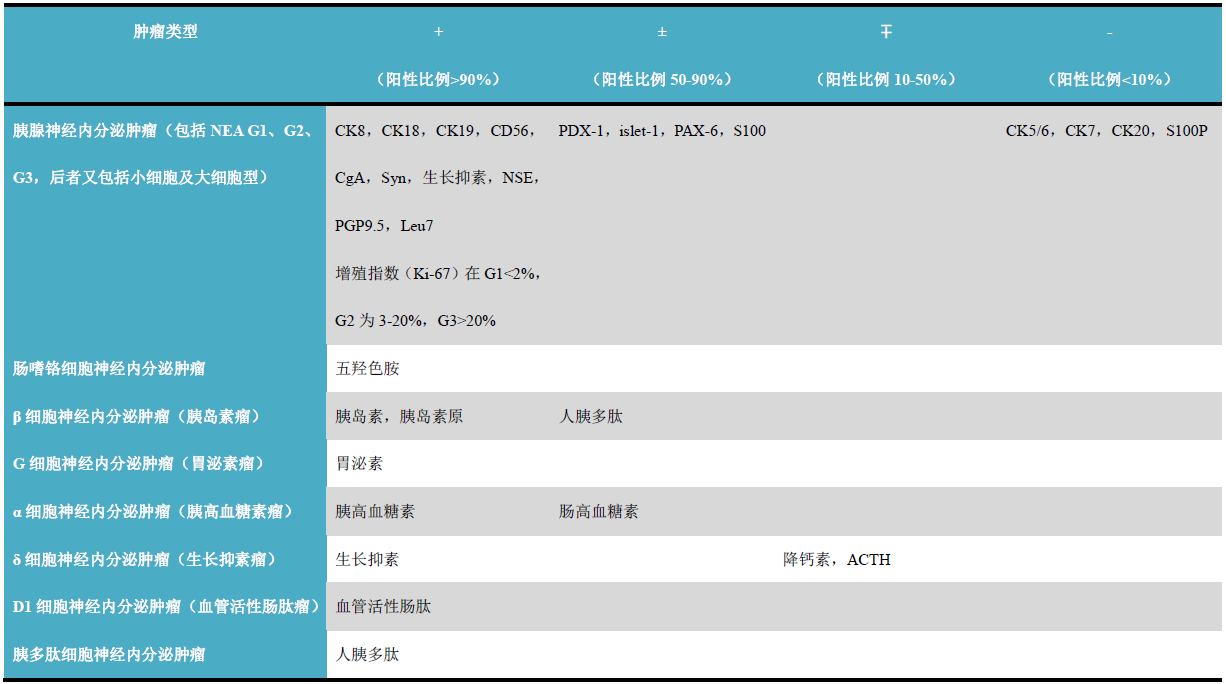
(Click to view larger image)
Common Immunohistochemical Indicators for Pathological Diagnosis of Other Pancreatic Tumors
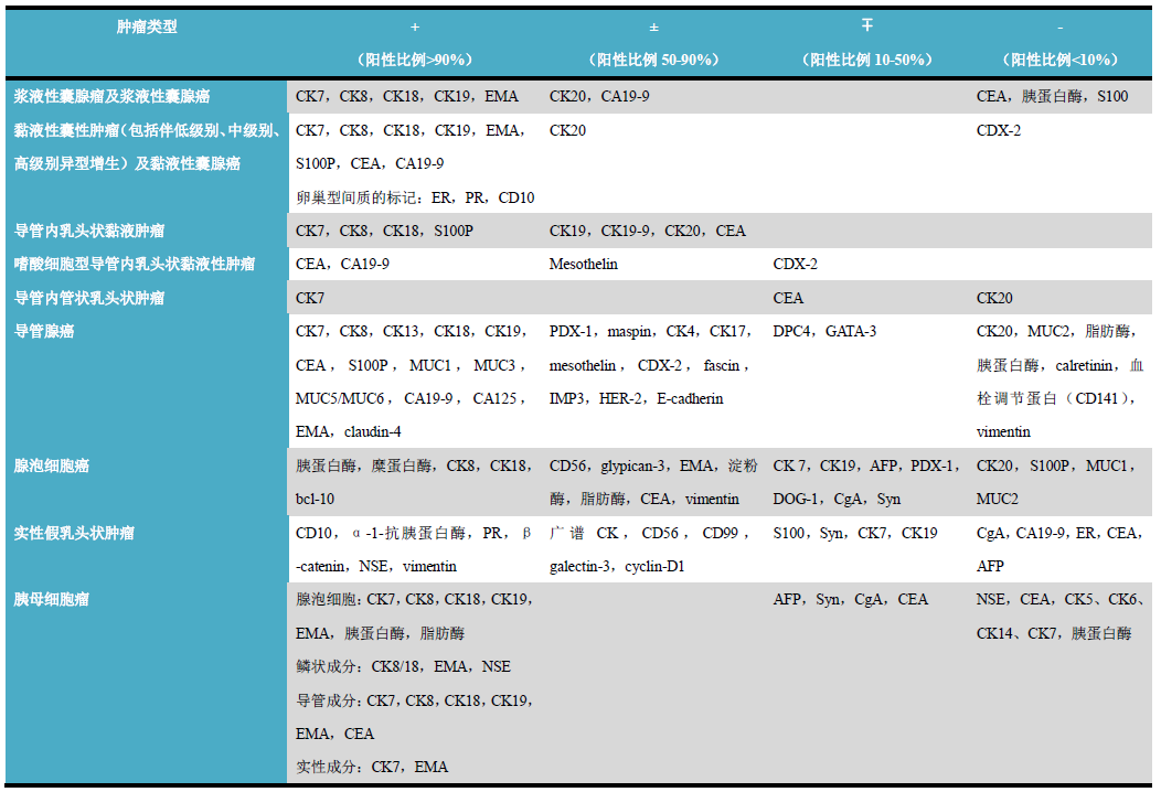
(Click to view larger image)
Detailed Explanation of Individual Indicators
CA19-9
Positive Expression Pattern: Cytoplasm
Recommended Positive Control Tissue: Pancreatic Tissue
PDX-1
Positive Expression Pattern: Nucleus
Recommended Positive Control Tissue: Pancreatic Tissue
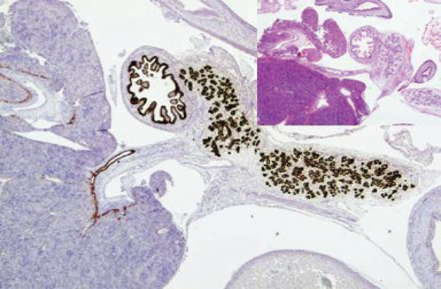
Figure 1. This case is a 12-week embryonic tissue. Immunohistochemistryshows PDX-1 expression in pancreatic ducts, duodenal mucosa, and bile duct mucosa.
S100P
Positive Expression Pattern: Cytoplasm/Nucleus
Recommended Positive Control Tissue: Pancreatic Cancer
Common Immunohistochemical Indicators for Differential Diagnosis of Pancreatitis and Pancreatic Cancer
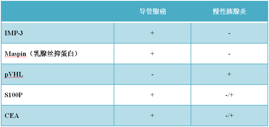
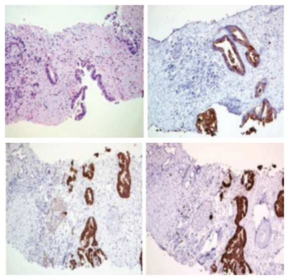
Figure 2. Core needle biopsy specimen of pancreatic ductal adenocarcinoma (top left). Immunohistochemistry shows malignant glands expressing CEA (top right) and IMP3 (bottom left), while these markers show very low positivity in islet cells. Malignant glands also express S100P (bottom right).
PAX-6
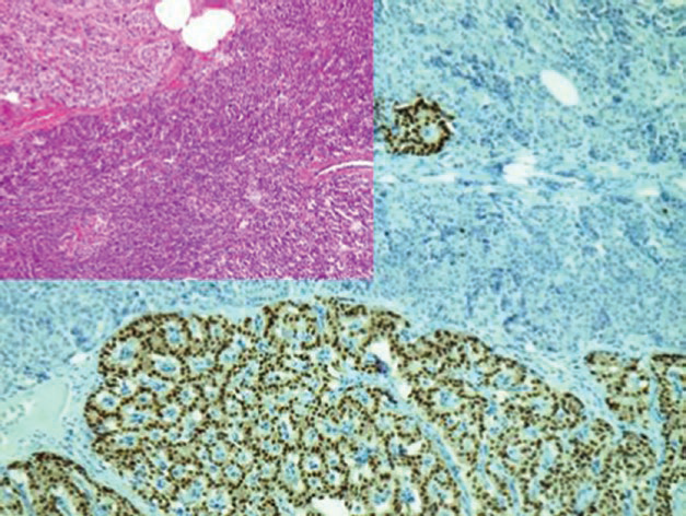
Figure 3. Pancreatic neuroendocrine tumor, G1. Immunohistochemistry shows tumor cells and endocrine cells in the islets expressing PAX-6.
1
Maimai: “CA19-9 is a tumor-associated carbohydrate antigen related to the Lewis A blood group antigen. Most gastrointestinal and pancreatic cancers express it positively, while tumors from other sites are less common. It is mainly used in the study of pancreatic cancer, colorectal adenocarcinoma, gastric cancer, and other tumors; it can also be used for the differential diagnosis of adenocarcinoma and mesothelioma. In pancreatic lesions,pancreatic cancer commonly shows positive expression of IMP3, but it is not expressed in benign pancreatic tumors, inflammatory pancreatic lesions, or normal pancreatic tissue, so it can be used for the differential diagnosis of pancreatic ductal adenocarcinoma. S100P is highly expressed in pancreatic ductal adenocarcinoma, while benign pancreatic ducts and acinar glands do not express it. Additionally,S100Pit is also one of the specific markers for urothelial carcinoma.”
|
Antibody Name |
Clone Number |
Positive Control |
Positive Location |
|
CA19-9 |
121SLE |
Pancreas, Colonic Adenocarcinoma |
Cytoplasm |
|
IMP3 |
EP286 |
Pancreatic Cancer, Colonic Adenocarcinoma |
Membrane/Cytoplasm |
|
S100P |
16/f5 |
Pancreatic Cancer, Placenta |
Nucleus/Cytoplasm |
For more information, please contact: 800-8581156 or 400-889-9853