Hotspot Focus

The 11th Issue of “Mai Mai” Pathology Weekly Reading Notes | Immunohistochemical Markers for Gastrointestinal Tumors (Part 2): Mesenchymal Tumors


Preface:


In the previous article, we mentioned that “gastrointestinal specimens occupy a significant portion of the daily work of pathologists.” We compiled and introducedknowledge of commonly used immunohistochemical markers in gastrointestinal tumors. In this issue, we will introducein gastrointestinal tumorsepithelial tumorsthe knowledge of commonly used immunohistochemical markers. In this issue, we will introducethe mesenchymal tumor section.


图片

Immunohistochemical Markers for Gastrointestinal Tumors: Mesenchymal Tumors

Table 1. Overview of Immunohistochemical Indicators for Gastrointestinal Mesenchymal Tumors 

图片

Remarks

  1. Gastrointestinal stromal tumors with epithelioid morphology are generally CD117 negative;

  2. In CD117-negative gastrointestinal stromal tumors,PDGFR-αpositive;

  3. Nuclear and cytoplasmic staining.


图片

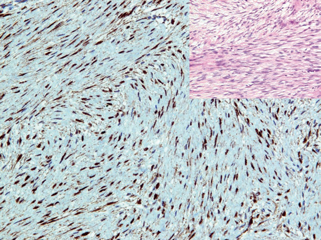
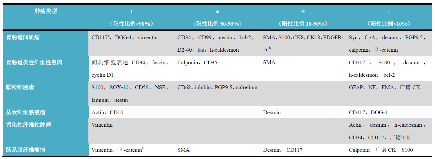
Figure 1. Mesenteric fibromatosis, β-cateninstrongly positive expression in the nucleus.


12


CD117

CD117is a tyrosine kinase growth factor receptorIIIfamily member, specifically composed of three parts: an extracellular domain, a transmembrane domain, and an intracellular kinase domain. Other members of this family includePDGFR-α, macrophage colony-stimulating factor,FMA-like tyrosine kinase3. Normally, CD117activation occurs after stem cell factor binding and is involved in the differentiation of hematopoietic cells, mast cells, germ cells, melanocytes (melanocytes), and intestinalCajalcells; therefore, in daily work, CD117expression is very broad and can be used as a diagnostic marker for various tumors, such as seminoma, mast cell tumor, chronic and acute myeloid leukemia, thymoma, adenoid cystic carcinoma, some acuteTlymphoblastic leukemia, and various malignant melanomas.
CD117expression can be seen in90%or more of gastrointestinal stromal tumors; approximately80%of gastrointestinal stromal tumors havec-Kitgene single or multiple activating mutations, mainly located in11exon, and less commonly seen in9exon,13exon,17exon. CD34and DOG1co-expression is also a characteristic immunohistochemical feature of this tumor.5-8%of gastrointestinal stromal tumors also havePDGFR-αgene mutations, and generally CD117negative; these tumors often exhibit epithelioid morphology, and immunohistochemistry is mostlyPDGFR-αand/or DOG1positive.

图片

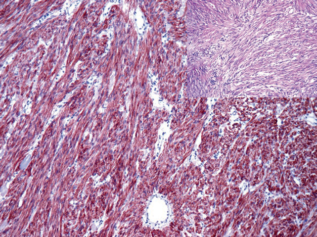
Figure 2. Gastrointestinal stromal tumor,CD117strongly positive.


Positive expression pattern: cell membrane/cytoplasm

Recommended positive control tissue: brain tissue


2


DOG1

DOG1is a transmembrane chloride channel protein, highly expressed in gastrointestinalCajalcells. This marker is highly specific for gastrointestinal stromal tumors, with a positive rate reaching90%or more. However, DOG1and CD117expression in gastrointestinal stromal tumors is highly consistent, but the former is not expressed in seminoma, myeloid-related tumors, or mast cell tumors. DOG1also helps differentiate salivary acinic cell carcinoma from other morphologically similar adenocarcinomas, but cannot be used to distinguish from bile ductpancreatic adenocarcinoma.

图片

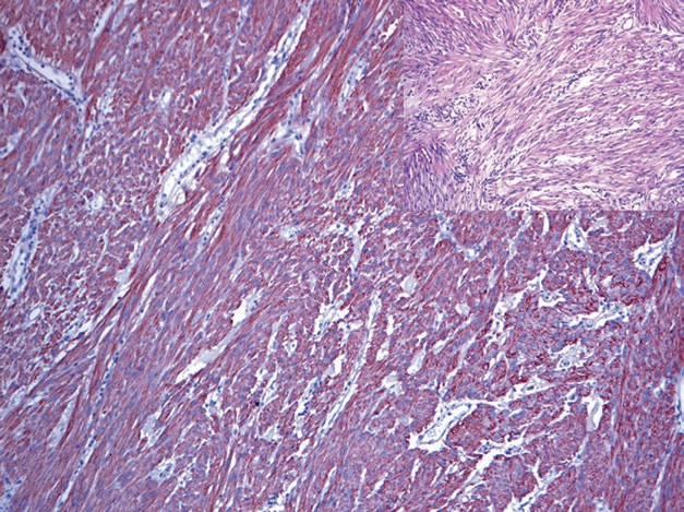
 Figure 3. Gastrointestinal stromal tumor, DOG1strongly positive.


up to50%of gastrointestinal intramural leiomyomas show low-level expression of DOG1, but these tumors generally strongly express Actinh-caldesmo, which can be used for differentiation.


Positive expression pattern: cell membrane/cytoplasm

Recommended positive control tissue: gastrointestinal stromal tumor


3


PDGFR-α

PDGFR-α is a tyrosine kinase receptor and also a member of theIIItype tyrosine kinase receptor family, related to embryonic development and immune responses in different tissues. As mentioned earlier,PDGFR-αis an important marker in CD117negative gastrointestinal stromal tumors, because CD117negative gastrointestinal stromal tumors havePDGFR-αgene activating mutations, while CD117positive gastrointestinal stromal tumors generally do not havePDGFR-αexpression.
PDGFR-α immunohistochemical results interpretation must consider that some desmoid tumors (desmoid tumors) also show positive expression of this marker. Normally,PDGFR-α is also expressed in ganglion cells, Schwann cells, thyroid follicular cells, and spermatogonia.


4


CD34

CD34is a cell surface adhesion glycoprotein, generally considered a vascular endothelial marker. CD34can be expressed in most gastrointestinal stromal tumors, but it is not specific, so it must be used in combination with DOG1and CD117. For gastrointestinal stromal tumors, CD34can also be expressed in stromal cells of gastrointestinal inflammatory fibroid polyps (inflammatory fibroid polyp).

图片

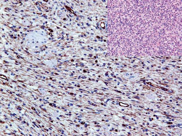
Figure 4. Stromal cells in gastrointestinal inflammatory fibroid polyps expressCD34

1


Maimai: “CD34 is mainly expressed in immature hematopoietic stem cells, myeloid cells, and vascular endothelial cells. It can identify endothelial cell differentiation, but the sensitivity for identifying blood vessels is not related to tumor grade. Over 85% of angiosarcomas and Kaposi sarcomas show positive CD34 expression. It is used in combination with CD117 for the diagnosis of gastrointestinal stromal tumors. Combined with the above,DOG1 is selectively expressed in gastrointestinal stromal tumors (GIST). Compared to CD117, DOG1 is a more sensitive and specific marker for GIST, regardless of whether the GIST has C-kit gene mutations or PDGFR-α gene mutations.”

Antibody Name

Clone Number

Positive Control

Positive Location

CD34

QBEnd/10

Hemangioma, liver

Cell membrane/cytoplasm

CD117

YR145

Gastrointestinal stromal tumor, seminoma

Cell membrane/cytoplasm

DOG1*

MX067

Gastrointestinal stromal tumor

Cell membrane/cytoplasm

DOG1

SP31

Gastrointestinal stromal tumor Cell membrane/cytoplasm

*Marked as Maxin clone product


For more information, please contact: 800-8581156 or 400-889-9853

发表回复

您的邮箱地址不会被公开。 必填项已用 * 标注