
Tenth Issue of “Mai Mai” Pathology Weekly Reading Notes | Immunohistochemical Markers for Gastrointestinal Tumors (Part 1): Epithelial Tumors
Preface:
Gastrointestinalspecimens occupy a significant portion of the daily work of pathologists. Undoubtedly, immunohistochemistry plays a very important role in their diagnosis and differential diagnosis. To facilitate a more systematic and comprehensive understanding for fellow pathologistsof the knowledge regarding commonly used immunohistochemical markers for gastrointestinal tumors,we will introduce them in two parts: epithelial tumors and mesenchymal tumors.

Immunohistochemical Markers for Gastrointestinal Tumors: Epithelial Tumors
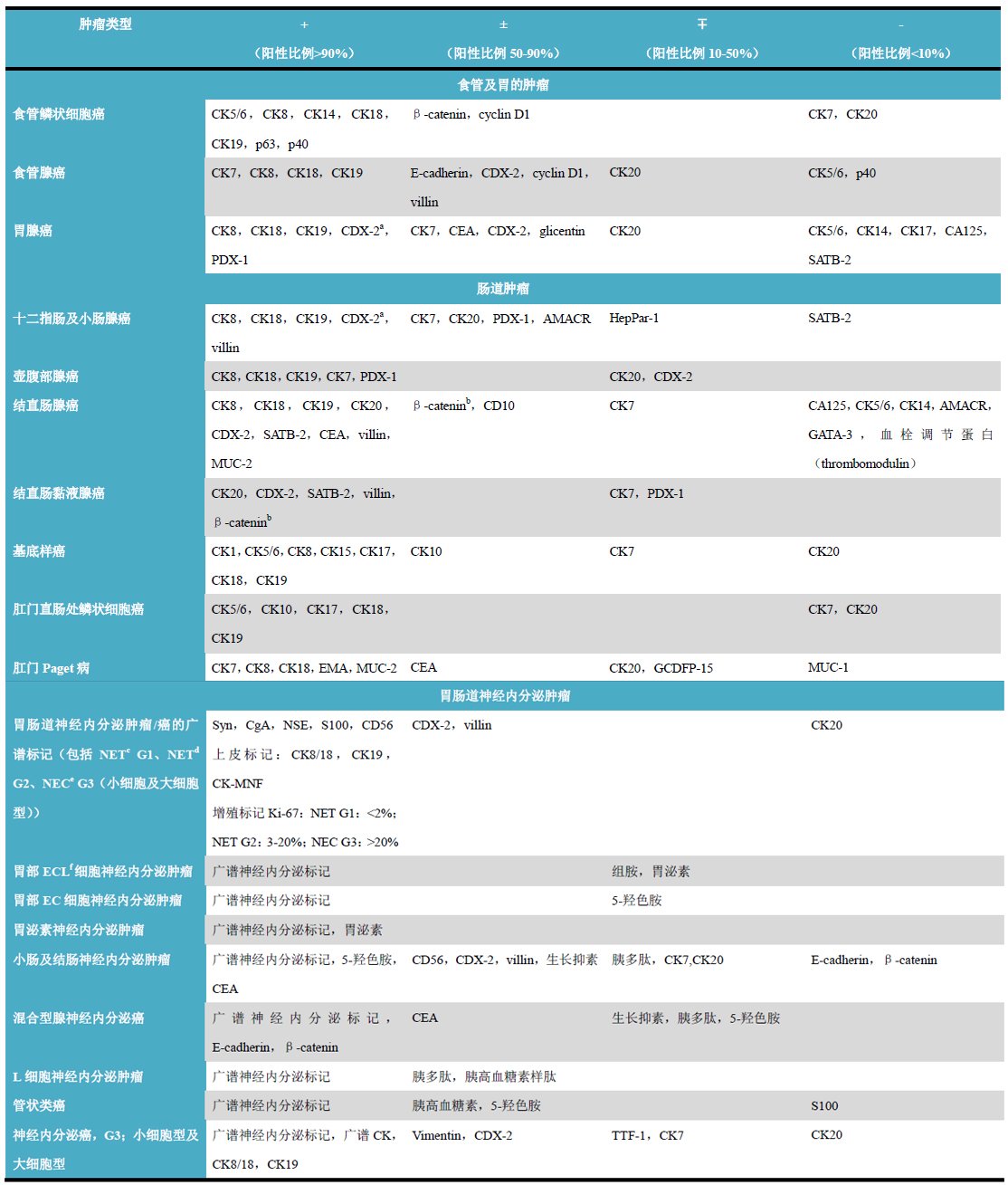
(Click to view larger image)
Remarks
-
The medullary type in adenocarcinoma is generally negative;
-
Nuclear staining;
-
Well-differentiated neuroendocrine tumor (carcinoid);
-
Well-differentiated neuroendocrine carcinoma (atypical carcinoid);
-
Poorly differentiated neuroendocrine carcinoma;
-
Enterochromaffin-like cells.
Detailed Explanation of Indicators
CDX-2
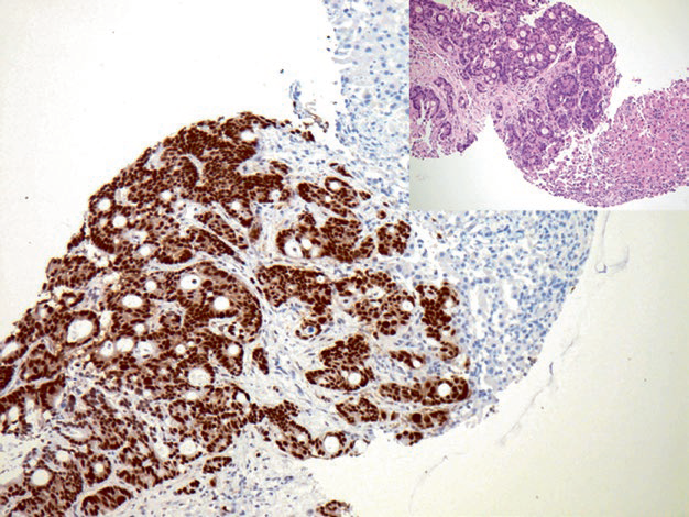
Figure 1. Metastatic colorectal adenocarcinoma, with strong nuclear positive expression of CDX-2 in tumor cells.
Positive expression pattern: Nuclear
Recommended positive control tissue: Appendix
SATB2
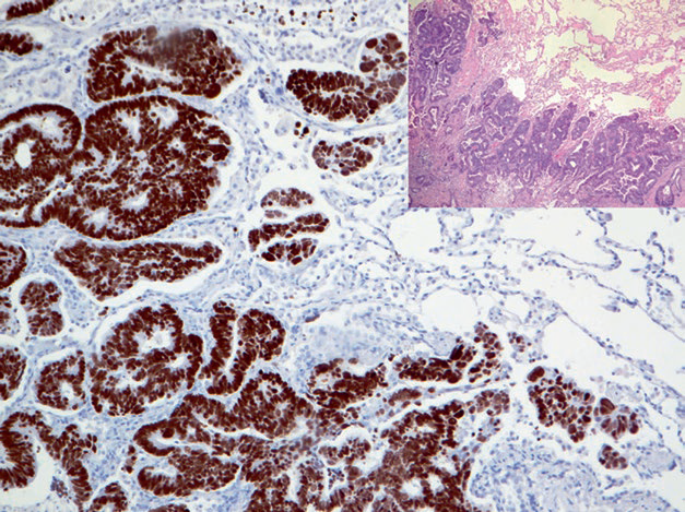
Figure 2. Rectal adenocarcinoma with lung metastasis, showing strong nuclear positive expression of SATB-2 in tumor cells.
Positive expression pattern: Nuclear
Recommended positive control tissue: Appendix
CDH17
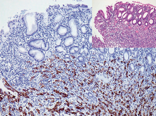
Figure 3. Positive CDH17 in gastric adenocarcinoma.
Positive expression pattern: Cell membrane and cytoplasm
Recommended positive control tissue: Appendix
Villin
1
Maimai: “CDX2 is an intestine-specific transcription factor expressed early in intestinal development, involved in regulating the proliferation and differentiation of intestinal epithelial cells, thus playing an important role in gastrointestinal epithelial metaplasia and tumorigenesis. Normally, this marker is expressed in the villi and crypt surfaces of intestinal epithelium, from duodenal to rectal epithelial cells. Used in combination with Villin, it can differentiate colorectal adenocarcinoma from metastatic adenocarcinoma. CDX2 expression is closely related to the occurrence of gastric cancer, detectable in 74% of gastric cancers. It is important to note that loss of CDX2 promotes the development of sporadic colon cancer, as mentioned in this article, medullary carcinoma in colon adenocarcinoma does not express this marker. SATB2 is mainly used for the diagnosis of osteosarcoma and its differential diagnosis from other non-osteogenic sarcomas. Additionally, this marker is highly expressed in colorectal cancer, while other tumors such as pancreatic, gastric, gallbladder, ovarian, cervical, and endometrial adenocarcinomas rarely express it or are negative, thus it can also serve as a sensitive and specific marker for lower gastrointestinal tumors (e.g., colorectal cancer) when used in combination with CK7 and CK20.”
|
Antibody Name |
Clone Number |
Positive Control |
Positive Location |
|
CDX-2* |
MX024 |
Appendix, colon adenocarcinoma |
Nucleus |
|
CDX-2 |
EPR2764Y |
Appendix, colon adenocarcinoma |
Nucleus |
|
SATB2 |
EP281 |
Colon, osteosarcoma |
Nucleus |
|
Villin* |
MX021 |
Colonadenocarcinoma, appendix | Cytoplasm |
|
Villin |
CWWB1 |
Colonadenocarcinoma, appendix |
Cytoplasm |
*Marked as Maxim clone products
For more information, please contact: 800-8581156 or 400-889-9853