Performance of Maxin Antibodies in NordiQC Evaluations
In 2022, in the external quality assessment activities organized by the NordiQC Immunohistochemistry Quality Control Center, Maxin antibodies performed excellently as always,achieved very encouraging results.Run66, Run B34, Run H22, and Run C12It is worth mentioning thatthe antibodies participating in the evaluation,such as BSAP, SYP, SMH, Napsin A, CD10, HER2, ER, PD-L1, are all independently developed by Maxin, China-originated “clones”.At the same time,。
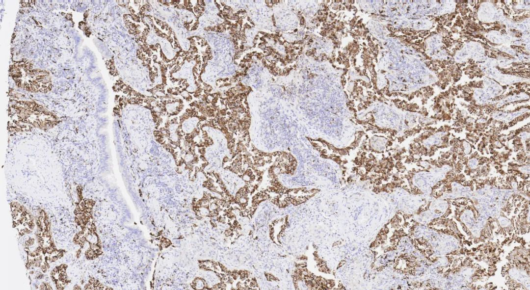
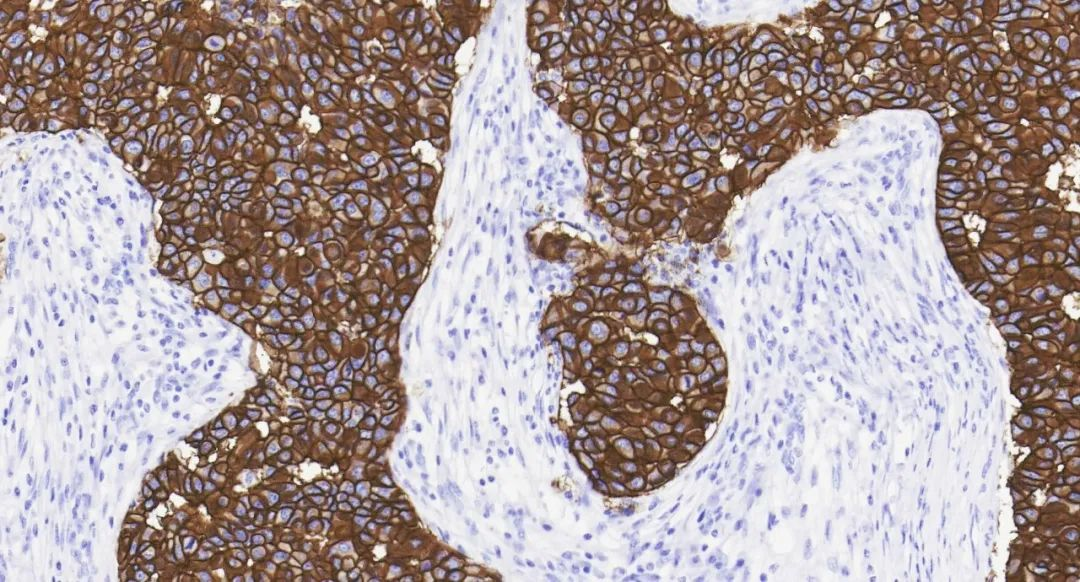
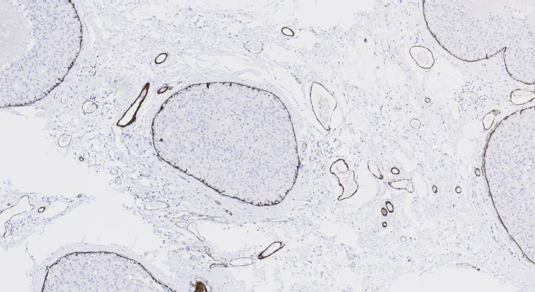
among the many global participants in NordiQC’s external quality assessment activities,Maxin is alsoone of the Chinese biopharmaceutical companiesFigure 1. Overview of Maxin Antibody Evaluation Resultswith the highest total number of completely independently developed antibodies and the most rated as excellent (Optimal).Napsin A (Aspartic proteinase napsin, Aspartic proteinase A) is mainly expressed in the lungs and kidneys, with low expression in the spleen. Its sensitivity in lung adenocarcinoma is significantly higher than that of SP-A and SP-B, and it has extremely high specificity. In some poorly differentiated lung adenocarcinoma cases where TTF-1 is negative, Napsin A is usually positively expressed. Therefore, it is recommended to be used in combination with TTF-1 for distinguishing primary lung adenocarcinoma from adenocarcinomas originating from other tissues and organs. It can also be used in combination with HNF1β for the diagnosis of ovarian clear cell carcinoma.。
321 laboratories participated, with a pass rate of 83% (excellent rate of 46%). Compared to the three previous evaluation results by NordiQC, the pass rate has improved this time.
In this round of NordiQC evaluation, a total of
Lung adenocarcinomaFigure 2.
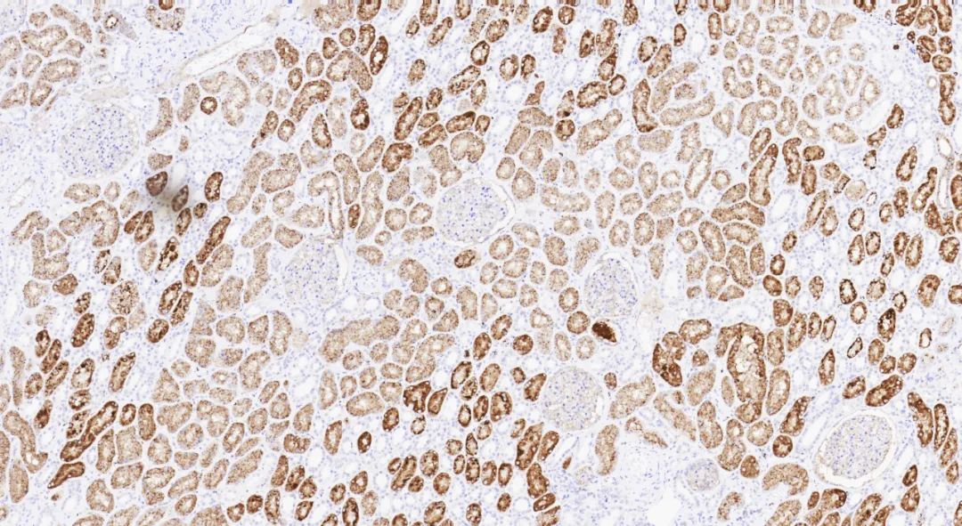
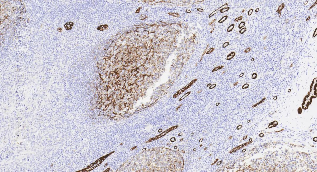
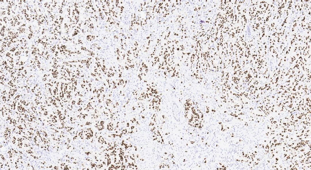
Napsin A (MX015) staining imageFigure 3. KidneySynaptophysin is mainly present in the presynaptic vesicle membranes of neurons, expressed in adrenal medullary cells, pancreatic islet cells, thyroid parafollicular C cells, parathyroid chief cells, skin, anterior pituitary cells, and neuroendocrine cells. It is primarily used for the diagnosis and differential diagnosis of pheochromocytoma, ganglioneuroma, paraganglioma, and tumors of the APUD system.
390 laboratories participated, with a pass rate of 70% (excellent rateSynaptophysin is mainly present in the presynaptic vesicle membranes of neurons, expressed in adrenal medullary cells, pancreatic islet cells, thyroid parafollicular C cells, parathyroid chief cells, skin, anterior pituitary cells, and neuroendocrine cells. It is primarily used for the diagnosis and differential diagnosis of pheochromocytoma, ganglioneuroma, paraganglioma, and tumors of the APUD system.
This round of evaluation is
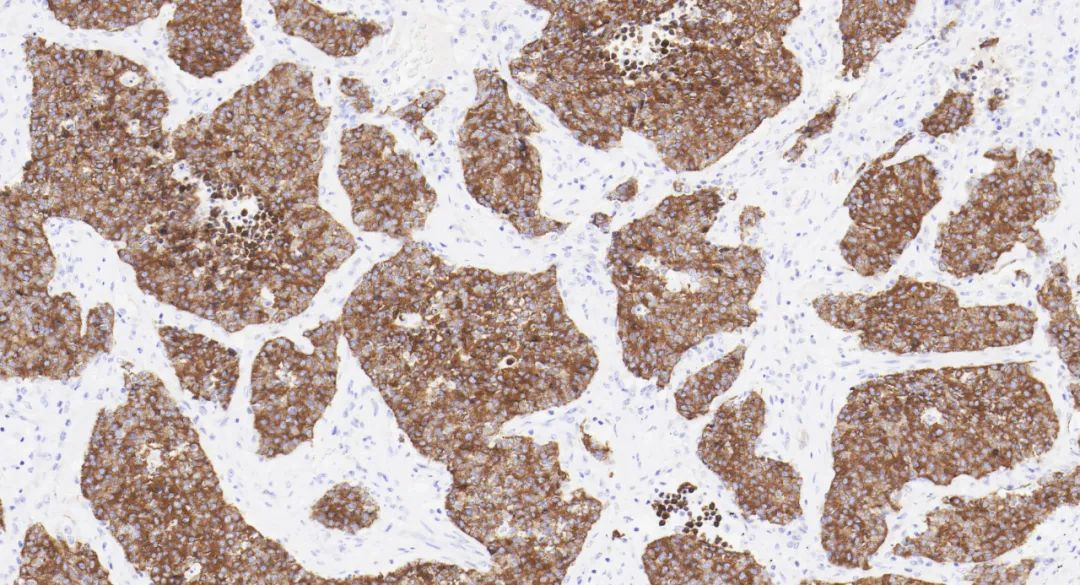
SYP, with a total ofNeuroendocrine tumorSYP (MX038) staining imageFigure 4.Figure 5.the seventh assessment by NordiQC forColon47%)。
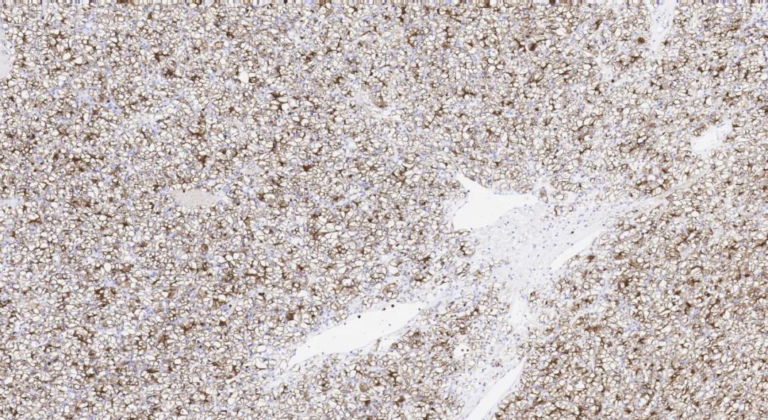
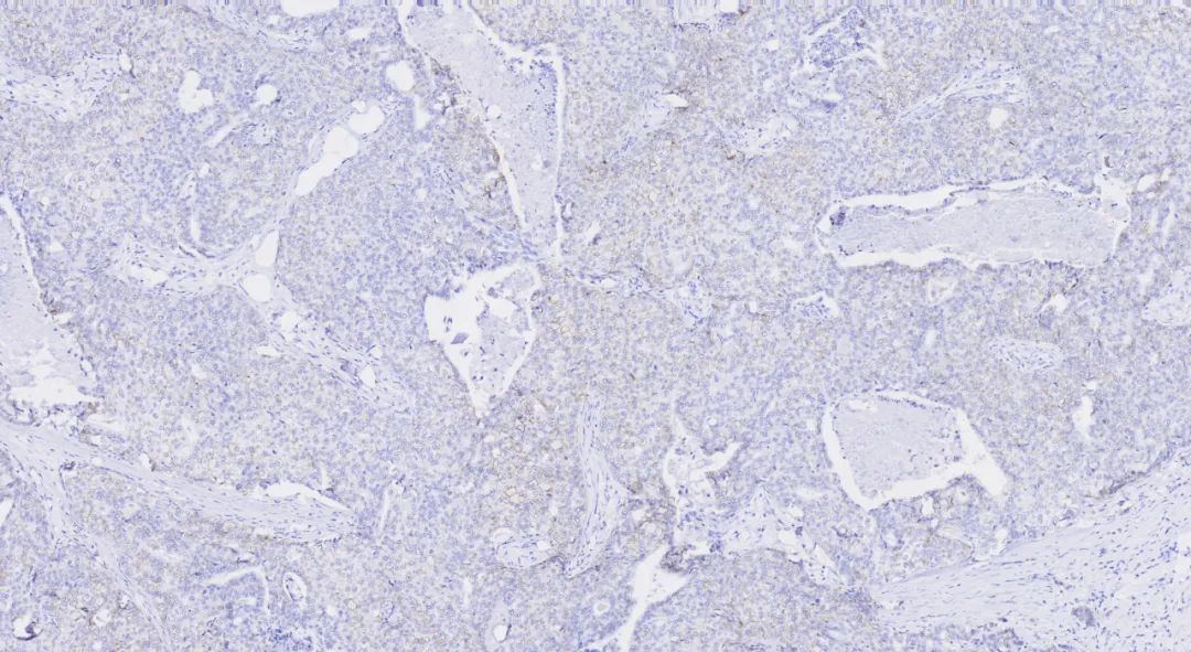
SMH, also known as Myosin (Smooth Muscle) (), is mainly expressed in vascular and visceral smooth muscle cells as well as myoepithelial cells, with occasional expression in myofibroblasts. It aids in the diagnosis and classification of mesenchymal tumors and, and can also be used for detecting breast myoepithelial cells.
Smooth muscle myosin heavy chainBreast, and can also be used for detecting breast myoepithelial cells.
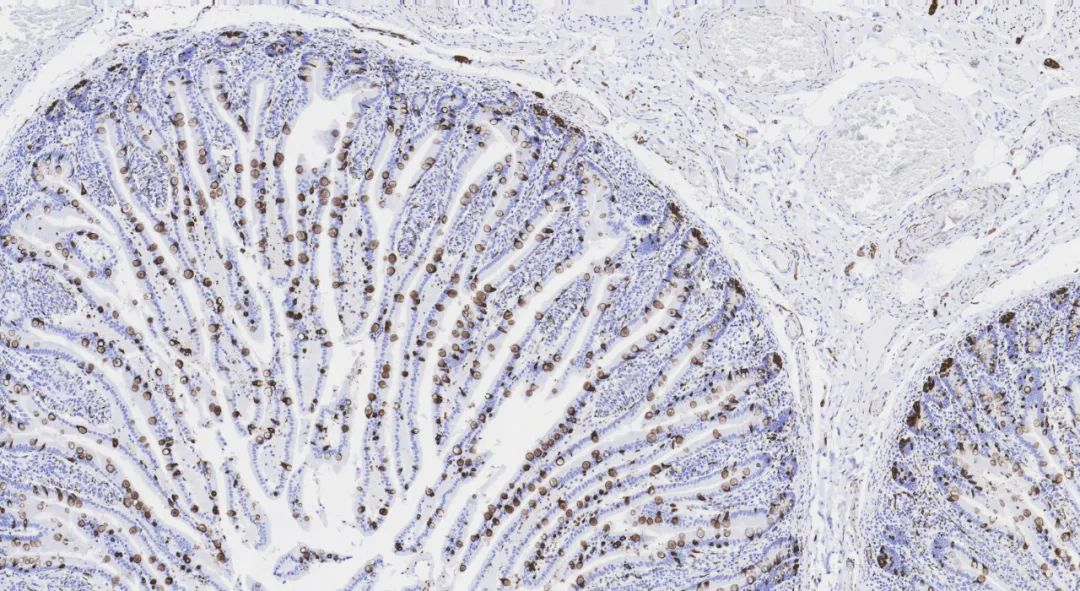
carcinoma in situ and invasive carcinomaIn this round of evaluation, a total of152 laboratories participated, with a pass rate of 81% (excellent rate wasIt should be noted that in the past, SMH was limited by antigen retrieval conditions (requiring58%). The focus of the evaluation was on the diagnostic use of SMH in breast samples to distinguish benign and pre-lesions from invasive carcinoma.
Figure 6.trypsin + citric acid high pressure), resulting in poor staining performance on automated platforms. However, Maxin’s SMH (MX109) antibody can now achieve excellent machine staining results under EDTA retrieval conditions.Breast carcinoma in situDual retrieval:
SMH (MX109) staining imageTonsilFigure 7.
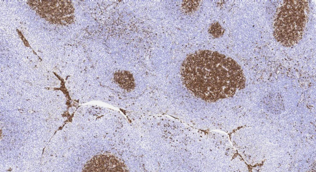
CD10, also known as CALLA (Common acute lymphocyte leukemia antigen), is a glycoprotein with a molecular weight of 100kDa, serving as a marker for follicular center cells. It can be used for the differential diagnosis of follicular lymphoma versus other low-grade malignant small B-cell lymphomas, renal clear cell carcinoma versus clear cell carcinomas from other origins, and endometrial stromal sarcoma versus uterine leiomyosarcoma, among other tumors.393 laboratories participated, but the pass rate was relatively low at 64% (This round of evaluation attracted
Maxin CD10 (MX002)Compared to foreign clones, it can achieve ideal staining results using a one-step method. This antibody has also received high praise and recognition from domestic and international customers due to its excellent performance in previous evaluation activities.This round of evaluation attracted
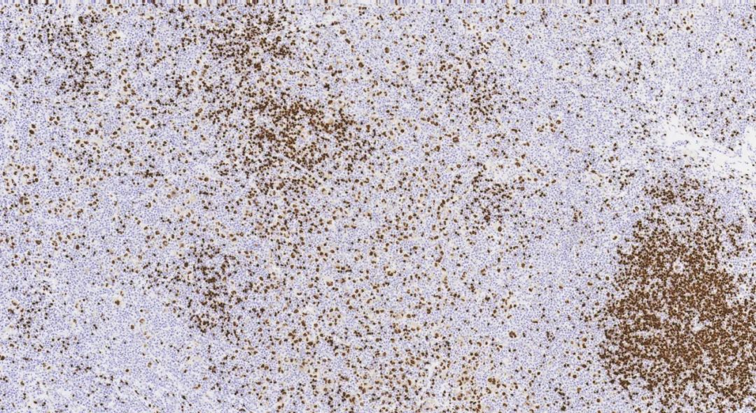
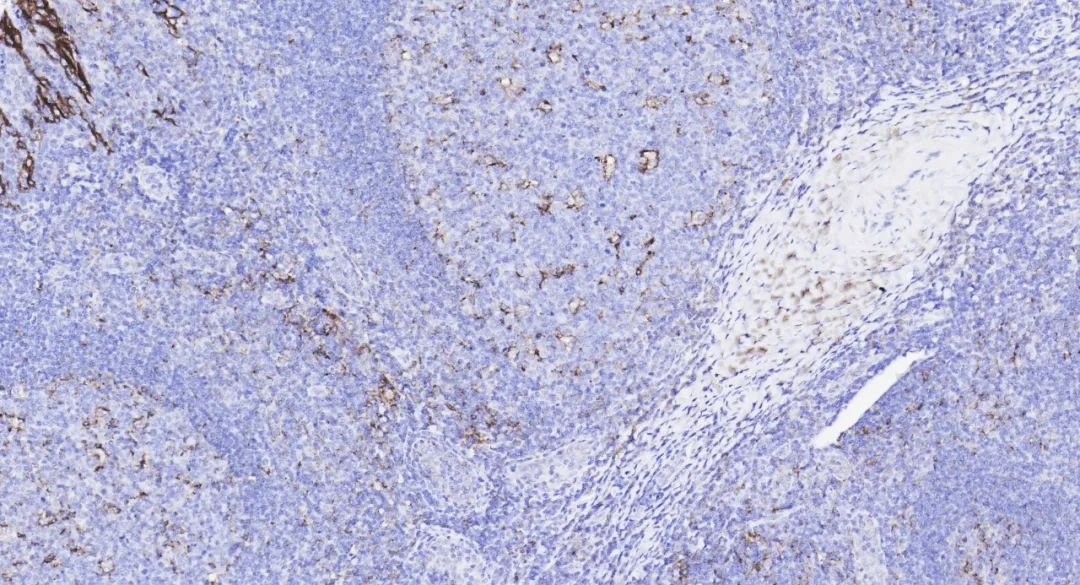
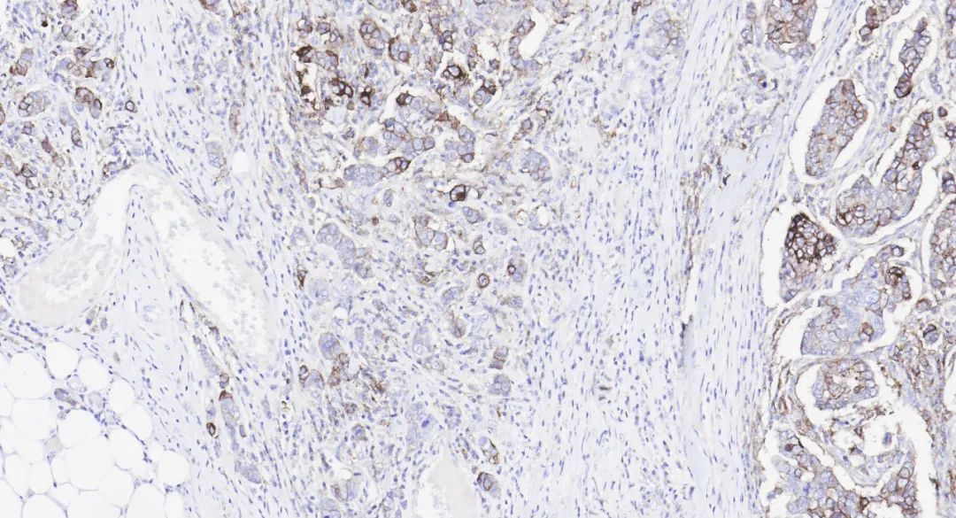
CD10 (MX002) staining imageRenal clear cell carcinomaBreast carcinoma in situ37%)。Figure 9.BSAP, also known as Pax-5 (Paired box gene 5), is a member of the Pax nuclear transcription factor family, located on chromosome 9p13, encoding the B-cell lineage-specific activator protein (BSAP). This protein plays an important role in the development of B cells and neurons, mainly expressed in the nuclei of pre-B cells and mature B cells, but not in plasma cells. It is primarily used for the diagnosis of B cells and tumors derived from them.
259 laboratories participated, with a relatively high pass rate of 86% (In this round of evaluation, a total ofMaxin BSAP (MX017) antibody performed excellently, receiving an excellent rating itself. At the same time, multiple
domestic and internationalCompared to foreign clones, it can achieve ideal staining results using a one-step method. This antibody has also received high praise and recognition from domestic and international customers due to its excellent performance in previous evaluation activities.Maxin BSAP (MX017) antibody performed excellently, receiving an excellent rating itself. At the same time, multiple
units using Maxin clones in the evaluation also received excellent ratings.
Hodgkin lymphomaFigure 10.Breast carcinoma in situ52%)。BSAP (MX017) staining imageFigure 11.HER2 (Human epidermal growth factor receptor 2), also known as C-erbB-2, is a proto-oncogene. Its activation can directly lead to malignant transformation or increased malignant potential in some human cells. Studies have shown that amplification of the HER2 gene contributes to the development of certain tumors such as breast cancer, gastrointestinal cancer, ovarian cancer, and endometrial cancer. In recent years, numerous new drugs have emerged in the field of anti-HER2 targeted therapy, especially with the advent of new ADC drugs, highlighting the significance of immunohistochemistry as a companion diagnostic.In thisThe pass rate for 392 laboratories was 84% (
round of evaluation, globally,excellent rateFigure 12.
BreastCompared to foreign clones, it can achieve ideal staining results using a one-step method. This antibody has also received high praise and recognition from domestic and international customers due to its excellent performance in previous evaluation activities.Figure 12.
HER2 (MXR011) staining imageFigure 13.invasiveInvasive breast carcinomaColon70%)。
, non-special) staining imagetypeER (Estrogen receptor) is
one of the members of the steroid hormone receptor protein superfamily. Clinical data indicate that the level of ER in breast cancer tissue is an important parameter for tumor prognosis and endocrine therapy. Breast cancer patients with positive ER expression often respond well to hormone therapy and have a favorable prognosis.406 laboratories participated, achievinga pass rate (In thisround, the global participantsHER2(MXR011a high 93%
Figure 14.ER (MXR034) staining imageInvasive breast carcinoma, non-special type
232 laboratories participated, with a qualification rate of 85%.This round of evaluation is the second assessment by NordiQC in 2022 for PD-L1 based on TPS/CPS status,Figure 15.with a total ofPD-L1 (Programmed cell death ligand 1), also known as CD274 or B7-H1, is a type I transmembrane protein with a molecular weight of 40kDa, involved in cellular regulation and immune responses. Studies show that the expression level of PD-L1 in tumor tissue samples is related to the efficacy of PD-1/PD-L1 inhibitor therapy. It can serve as a companion diagnostic to guide treatment decisions with PD-1/PD-L1 monoclonal antibodies and as a supplementary diagnostic to help identify potential beneficiaries of immunotherapy.Invasive breast carcinomaColon63%)。
Maxin(MX070C) antibody has consistently achieved excellent (Optimal) results since participating inquality control activities in 2019, demonstrating good performance advantages.
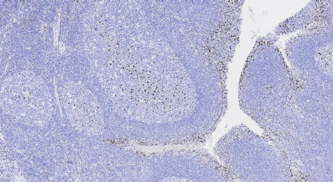
Figure 16.Compared to foreign clones, it can achieve ideal staining results using a one-step method. This antibody has also received high praise and recognition from domestic and international customers due to its excellent performance in previous evaluation activities.quality control activities in 2019, demonstrating good performance advantages.
Figure 17.TEXT_117For more information, please contact: 800-8581156 or 400-889-9853PD-L1 (MX070C) staining imageTEXT_118PD-L1TEXT_119NordiQCTEXT_120
TEXT_124Compared to foreign clones, it can achieve ideal staining results using a one-step method. This antibody has also received high praise and recognition from domestic and international customers due to its excellent performance in previous evaluation activities.TEXT_123
TEXT_125