GATA3 binding protein, commonly abbreviated as GATA3, is a zinc finger transcription factor in the GATA family. It plays an important role in promoting and guiding cell proliferation, development, and differentiation in various tissues and cell types. In normal tissues, GATA3 is often expressed in the nuclei of cells in the breast, skin, kidney, urothelium, and large vessel epithelium. It shows weak nuclear expression in interfollicular T cells of lymph nodes. It is not expressed in the thyroid, respiratory tract, alveolar epithelium, stromal cells, hepatocytes, ovary, endometrium, blood vessels, and other soft tissues, thus it can be used for internal quality control in tissues. In tumor tissues, GATA3 shows positive expression in primary and metastatic ductal carcinoma and lobular carcinoma. It is also used to differentiate urothelial carcinoma from prostate cancer.
Among mesotheliomas, malignant pleural mesothelioma is the most common type (81%). Besides the pleura, mesothelioma can also occur in other sites, such as the peritoneum (9%), pericardium, and tunica vaginalis testis. The World Health Organization classifies the histological subtypes of malignant mesothelioma (DMM) into epithelioid (most common), sarcomatoid, and biphasic. Due to the multipotent differentiation function of mesothelial cells, malignant mesothelioma cells exhibit diverse morphologies, making it difficult to distinguish between adenocarcinoma and reactive mesothelioma, sarcomatoid mesothelioma and sarcomatoid carcinoma. This is particularly true for pulmonary sarcomatoid carcinoma, which consists of atypical spindle cells and sometimes mixes sarcomatoid carcinoma with squamous or adenoid carcinoma. Sarcomatoid mesothelioma is a poorly differentiated malignant tumor arising from mesothelial cells and also contains atypical spindle cell components arranged in bundles, nodules, or occasionally haphazard patterns. Both tumors exhibit high-grade histological features without specific morphological characteristics, making differential diagnosis exceptionally challenging.
Extensive studies have found that the expression rate of GATA3 is relatively high in reactive mesothelioma and malignant mesothelioma (58%). In contrast, it is rarely expressed in lung cancer and squamous cell carcinoma (0-8%). GATA3 is usually positive in the glandular part of biphasic synovial sarcoma and positive in 20% of epithelioid sarcomas. In the vast majority of other sarcomas, the GATA3 positivity rate ranges from 0% to 10%. Based on the above, this study aims to investigate whether diffuse sarcomatoid malignant mesothelioma (SMM/DMM) expresses GATA3. If GATA3 is expressed, it could be used to differentiate SMM/DMM mesothelioma from pulmonary sarcomatoid carcinoma.
In this study, 19 cases of SMM/DMM mesothelioma and 13 cases of pulmonary sarcomatoid carcinoma were collected for GATA3 immunohistochemical experiments. The staining results were scored based on staining intensity and extent. The diffuse dispersion of tumor cells was graded from 0 to 3: <1% as grade 0, 1%-25% as grade 1, 25%-50% as grade 2, >50% as grade 3. Staining intensity was scored from 0 to 3 as follows: 0 for none, 1 for weak, 2 for moderate, and 3 for strong staining. The two scores were summed to obtain the final score, with a maximum score of 6.
The results of GATA3 immunohistochemical staining are shown in Table 1. All cases used inflammatory cell nuclear staining as an internal control. All 19 cases of SMM/DMM mesothelioma showed diffuse moderate to strong positive staining (mean ± standard deviation for 19 cases: 5.4 ± 0.9, see figure below). Among the 13 cases of pulmonary sarcomatoid carcinoma, most showed no staining except for positive controls. Two cases showed GATA3 positive staining (15%), which was only focal and weak (both cases scored 2 points; mean ± standard deviation for 13 cases: 0.4 ± 0.8, see figure below). The study results showed a significant difference in scores between the two groups (p < 0.001). Additionally, there was no clear correlation between GATA3 staining intensity and extent and traditional mesothelioma markers.
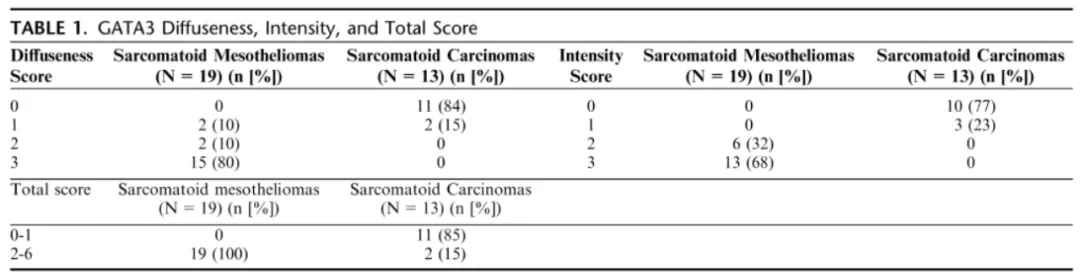
SMM/DMM mesothelioma (A: HE staining, B: GATA3 staining, showing strong diffuse staining);
Pulmonary sarcomatoid carcinoma (C: HE staining, D: GATA3 staining result, with inflammatory cells serving as strong positive internal control, tumor cells negative).
The study found that using GATA3 immunohistochemical detection for differentiating SMM/DMM mesothelioma from pulmonary sarcomatoid carcinoma had a sensitivity of 100% and a specificity of 85%. Therefore, this study indicates that GATA3 is a sensitive and reasonable immunomarker in the diagnosis of mesothelioma for distinguishing sarcomatoid mesothelioma from sarcomatoid carcinoma.
Two points should be noted in clinical use: First, when tumor cells are not completely stained, results with inflammatory cell nuclear staining are reliable. Second, this result is not absolute because weak focal positive staining is occasionally seen in sarcomatoid carcinoma, requiring other indicators for auxiliary differentiation. Positive staining in inflammatory cells can serve as an internal quality control; if tumor cell staining intensity is weaker than the internal control, it tends to favor sarcomatoid carcinoma.
Related Antibodies from Maxin
|
Antibody Name
|
Product Number
|
Clone Number
|
Positive Location
|
|
GATA3
|
MAB-0695
|
L50-823
|
Nuclear
|
*Marked as Maxin clone products
Source Literature:
1. Berg, K. B., & Churg, A. GATA3 Immunohistochemistry for Distinguishing Sarcomatoid and Desmoplastic Meso-thelioma From SarcomatoidCarcinoma of the Lung. The American Journal of Surgical Pathology, (2017). 41(9),1-221–1225.
