In tumor diagnosis, the expression of HNF1β has a certain correlation with the pathological types of liver cancer, specifically,it is highly expressed in intrahepatic cholangiocarcinoma and expressed in hepatocellular carcinoma.Germline mutations in HNF1β predispose to renal tumors, suggesting HNF1β as a tumor suppressor gene for renal chromophobe cell carcinoma.It can be considered that HNF1β mutations can lead to renal cysts in young people and diabetes in adulthood (MODY5).Additionally reported,HNF1βit is also a specific marker for the differential diagnosis of ovarian cancer.
Ovarian clear cell carcinoma
In 2003, Tsuchiya et al. used oligonucleotide array technology to study the differential gene expression between ovarian clear cell carcinoma and non-clear cell carcinoma. They found that HNF1β expression was significantly upregulated in clear cell carcinoma. RT-PCR, Western blot, and immunohistochemistry methods were used to confirm that HNF1β is a highly specific marker for ovarian clear cell carcinoma.
Nemegecova et al., while investigating genetic changes of HNF1-β in endometrial lesions, discovered truncated variants in ovarian clear cell carcinoma.This study identified four sequence variants and one missense mutation in ovarian clear cell carcinoma patients. Nonsense mutations lead to premature termination of translation, while missense variants have a disruptive effect on protein function.
Ka-to et al. analyzed the expression of HNF1β in 30 cases of ovarian clear cell tumors. The results showed that all cases expressed HNF1β, and among 12 cases with endometriosis epithelium, 9 also expressed HNF1β in the ectopic endometrial epithelium. However, other types of ovarian epithelial tumors (endometrioid, serous, mucinous, and Brenner) rarely expressed HNF1β. This study further confirmed that HNF1β is a good marker for ovarian clear cell tumors, including benign, borderline, and malignant clear cell tumors. Simultaneously, the expression of HNF1β in endometriosis indicates a potential for malignant transformation into ovarian clear cell carcinoma.
Xiong Kemei et al. used immunohistochemical EnVision method to detect 108 cases of epithelial carcinoma. The results showed that ovarian clear cell carcinoma expressed HNF1β to varying degrees, with a positive rate of 85.2% (27/27); the positive rate of HNF1β in ovarian high-grade serous carcinoma was 2.9% (4/35); in ovarian endometrioid adenocarcinoma, the positive rate was 23.8% (5/21); in ovarian mucinous carcinoma, HNF1β showed nuclear positivity, with a positive rate of 60.0% (10/10); Krukenberg tumors all showed varying degrees of nuclear positivity for HNF1β, with a positive rate of 53.8% (13/13); 2 cases of ovarian transitional cell carcinoma did not express HNF1β. The sensitivity and specificity of HNF1β for diagnosing ovarian clear cell carcinoma were 85.2% and 76.5%, respectively. The conclusion indicates that HNF1β is a highly sensitive marker for ovarian clear cell carcinoma. Diffuse strong positivity of HNF1β has high specificity for diagnosing ovarian clear cell carcinoma, especially in distinguishing it from ovarian serous carcinoma and endometrioid carcinoma with clear cell changes.
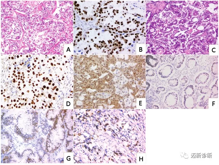
Figure 1. Expression of HNF1β in ovarian clear cell carcinoma; A. Tumor cells are arranged in nests, with clear cytoplasm and necrosis; B. HNF1β is localized in the nucleus, appearing yellow; C. Tumor cells are papillary, with hobnail cells and significant atypia; D. HNF1β is localized in the nucleus, appearing brown; E. HNF1β in serous carcinoma appears yellow and relatively vague; F. HNF1β in endometrioid carcinoma appears brown-yellow and relatively vague; G. HNF1β in mucinous carcinoma appears brown; H. HNF1β in metastatic Krukenberg tumor shows nuclear positivity.
Rougeont et al. reported in 2018 the expression of HNF1β in 45 cases of testicular and ovarian germ cell tumors, considering it a sensitive and reliable marker for diagnosing yolk sac tumors.
Gallo et al. used immunohistochemical staining to study the expression of HNF1β in tissue microarrays (TMA) of 601 testicular germ cell tumors, including seminoma, embryonal carcinoma, yolk sac tumor, choriocarcinoma, teratoma, in situ germ cell neoplasia, and normal tissues. They compared it with recommended markers for detecting yolk sac tumors, glypican-3 (GPC3) and alpha-fetoprotein (AFP). The study found that HNF1β (nuclear staining) exhibited a different staining pattern compared to GPC3 and AFP (cytoplasmic staining). The sensitivity and specificity of HNF1β were 85.4% and 96.5%, respectively; for GPC3, they were 83.3% and 90.7%; for AFP, they were 62.5% and 97.7%. HNF1β had higher sensitivity than AFP and higher specificity than GPC3. The nuclear expression pattern is easier to interpret; therefore, HNF1β is a specific marker for yolk sac tumors.
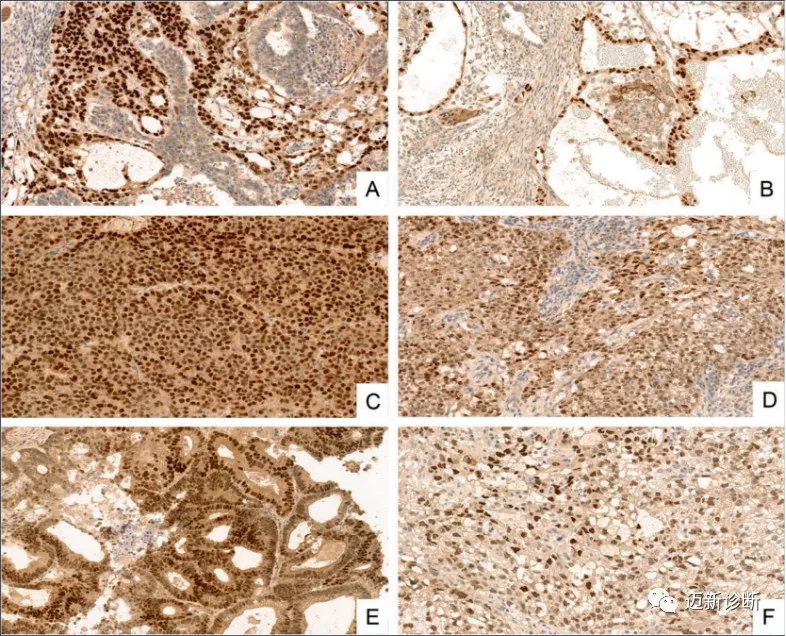
Figure 2. Expression patterns of HNF1β in different yolk sac tumors; A. Mixed embryonal; B. Macrovesicular; C. Hepatoid; D. Solid; E. Glandular; F. Microcystic/reticular.
Related antibodies from Maixin
|
Antibody Name
|
Product Number
|
Clone Number
|
Positive Location
|
|
HNF1β*
|
MAB-0884
|
MX092
|
Nucleus
|
*Marked asMaixin Clone Product
References:
[1] Alessandra Gallo, Christian Fankhauser, Thomas Hermanns et al. HNF1β is a sensitive and specific novel marker for yolk sac tumor:a tissue microarray analysis of 601 testicular germ cell tumors. Modern Pathology. 19 June 2020
[2] Xiong Kemei, Huang Wenbin, Wang Jinsong, et al. Diagnostic value of HNF-1β in ovarian clear cell carcinoma [J]. Journal of Clinical and Experimental Pathology, 2015(3):282-285.
[3] Wang Jinsong, Li Qing, Cheng Xue, et al. Value of combined detection of HNF-1β and Napsin A in the diagnosis of ovarian clear cell carcinoma [J]. Chinese Journal of Pathology, 2015,44 (12): 874-878.
