
The MUC5AC gene is located at 11p15.5 and is a secreted glycoprotein with mucosal protective functions. It participates in processes such as cell growth and differentiation, intercellular signal transduction, and intercellular adhesion, playing a protective and lubricating role in the mucosal epithelium. In recent years, numerous studies have shown that MUC5AC is abnormally expressed in diseases such as gastric cancer, colorectal cancer, lung cancer, and gallbladder lesions, and is involved in the occurrence and development of tumors, closely related to prognosis prediction.
During the occurrence and development of gastric cancer, metaplastic epithelium replaces the natural mucosa.As one of the important gastric-type secreted mucins, MUC5AC is a major component of mucus, the first protective barrier for mucosal epithelial colonization. In this process, its expression intensity decreases.
Jie Yang’s study downloaded MUC5AC expression data and survival information of 1140 gastric cancer patients from the CEO database. Analysis showed that MUC5AC expression in gastric tissue decreases with the development of intestinal metaplasia and is related to the stage of gastric cancer. The expression level of MUC5AC in advanced gastric cancer tissues is significantly lower than in early tissues and is associated with apoptosis pathways.This suggests that downregulation of MUC5AC expression is involved in the occurrence and development of gastric cancer and can serve as a new biomarker for gastric cancer prognosis.
Zhang Yuefan et al. conducted an IHC study on 78 gastric cancer tissues and adjacent normal tissues, finding that the positive expression rate of MUC5AC in gastric cancer tissues (48.72%, 38/78) was significantly lower than in adjacent normal tissues (92.31%, 72/78), mainly localized in the cytoplasm, appearing as yellow or brown-yellow granules (Figure 1, A, B). Patients with negative MUC5AC expression had significantly shorter survival times than those with positive expression (Figure 2), indicating that MUC5AC is an independent factor for predicting gastric cancer prognosis. High expression of MUC5AC suggests better differentiation of gastric cancer and possibly a better prognosis.
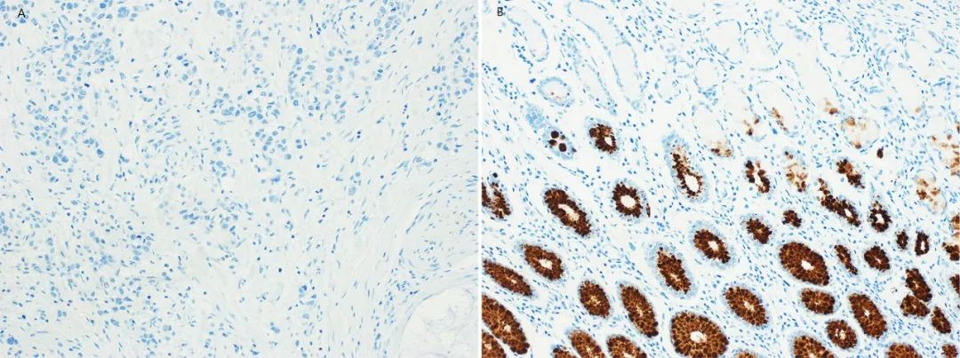
Figure 1. A: Negative expression of MUC5AC in gastric cancer; B: Expression of MUC5AC in adjacent normal tissue
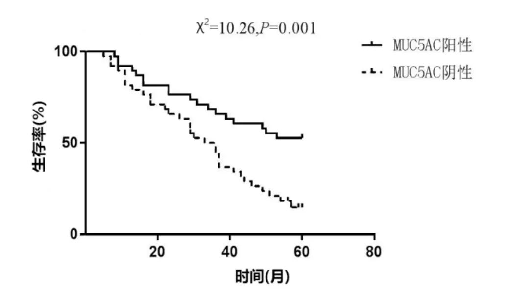
Figure 2. Survival curve of patients with MUC5AC expression in gastric cancer
The ability to secrete mucus is one of the important markers of colorectal cancer. Therefore, abnormal expression of mucins and changes in their glycosylation are closely related to the occurrence and development of colorectal cancer.
MUC5AC is typically expressed in the stomach and the inner lining of the trachea and bronchi, but not in normal intestinal tissue. Meriam HAZGUI et al. conducted an IHC study on 202 colorectal cancer cases in the Tunisian region, finding that MUC5AC expression was not significantly correlated with clinicopathological criteria such as stage, grade, and lymph node metastasis of colorectal cancer. However, the overall survival rate of positive patients was higher than that of negative patients.This suggests that the presence of MUC5AC may be a favorable prognostic factor for colorectal cancer and a new potential therapeutic target for colon cancer.
Hector Mesa et al. collected resection specimens of appendiceal and left/right colorectal tumors from 1999 to 2017. IHC studies showed that MUC5AC expression was higher in low-grade appendiceal mucinous neoplasms and high-grade mucinous adenocarcinomas compared to conventional colon adenocarcinomas and crypt cell adenocarcinomas. Compared to conventional colon adenocarcinomas, MUC5AC expression was more intense in high-grade mucinous adenocarcinomas. Additionally, the study found a significant correlation between MUC5AC expression and MMR deficiency, supporting serial reports that MUC5AC expression is higher in tumors arising via the serrated pathway (i.e., mucinous tumors with BRAF mutations and epigenetic MMR defects). The overall superior response of this molecular group to adjuvant therapy explains the favorable outcomes of colorectal cancer expressing MUC5AC.This indicates that MUC5AC expression in colorectal cancer is a potential marker for good prognosis and a therapeutic target.
Wu Qiang et al. conducted an IHC study on 42 cases of peripheral non-small cell lung cancer, finding that MUC5AC was positively correlated with lymph node metastasis of the tumor (p<0.05) and significantly positively correlated with tumor size, lobulation sign, spiculation sign, vascular convergence sign, and pleural invasion, indicating that MUC5AC plays an important role in the invasion and metastasis of lung cancer.
Yujie Dong et al. conducted an IHC study on samples from 90 patients with lung adenocarcinoma with mucin production (LAMP), finding different staining patterns of MUC5AC in different LAMP subtypes: positive staining along the apical surface of tumor cells in acinar-predominant adenocarcinoma (APA) and papillary-predominant adenocarcinoma (PPA), resembling an apocrine pattern; punctate distribution and focal staining in micropapillary-predominant adenocarcinoma (MPA) and solid-predominant adenocarcinoma (SPA); diffuse and dense staining in invasive mucinous adenocarcinoma (IMA) and colloid adenocarcinoma with goblet columnar cells (Figure 3 shows MUC5AC staining in APA, PPA, SPA, IMA).This indicates that LAMP has strong heterogeneity, and MUC5AC is an effective marker for distinguishing pure mucinous adenocarcinomas (including IMA and colloid adenocarcinoma, etc.) from non-pure mucinous adenocarcinomas (including APA, PPA, SPA, MPA, etc.).
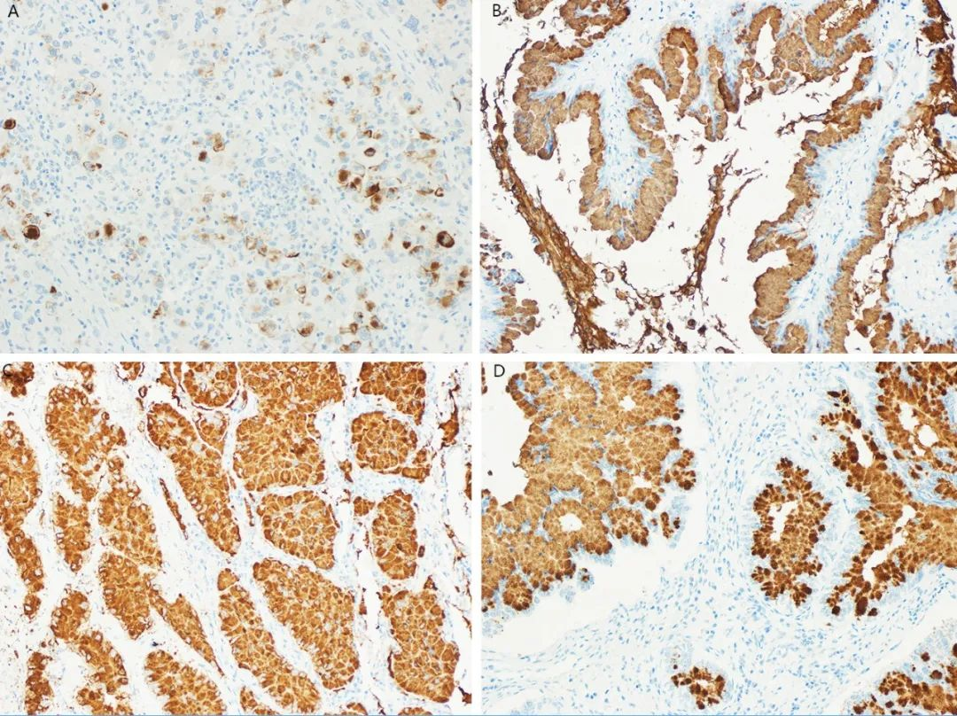
Figure 3. Staining patterns of MUC5AC in different LAMP subtypes (A: MUC5AC expression in acinar-predominant adenocarcinoma; B: MUC5AC expression in PPA; C: MUC5AC expression in SPA; D: MUC5AC expression in IMA)
Furthermore, during the multistage carcinogenesis of bronchial epithelial cells, the quantity of MUC5AC and mucin components increases. MUC5AC expression is more significant in IMA (P < 0.001) and in tumors with KRAS mutations (P = 0.005) and lacking TTF-1 expression (P < 0.001). Compared to pure mucinous subtypes, MUC5AC-positive cases in non-pure mucinous subtypes are more aggressive. Compared to MUC5AC-negative tumors, MUC5AC-positive tumors are positively correlated with poorer prognosis (Figure 4, P < 0.001). Multivariate survival analysisindicates thatMUC5AC is an independent prognostic factor for poor prognosis (Figure 4, P = 0.006) and may be a potential therapeutic target for this unique type of lung adenocarcinoma, which has few effective treatments.
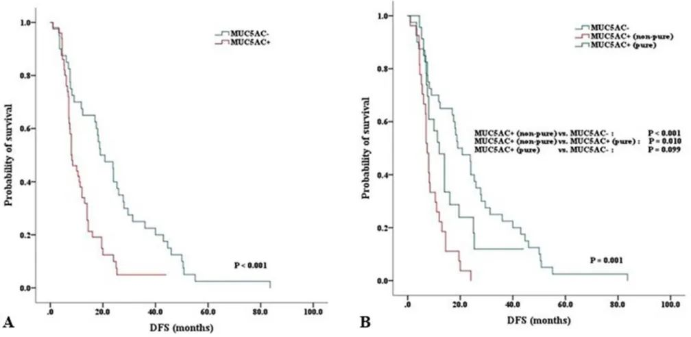
Figure 4. Kaplan-Meier survival curves for LAMP patients (A: Disease-free survival curve by MUC5AC expression status; B: Disease-free survival curve by pathological subtype and MUC5AC expression status)
Amit Bhoge et al. conducted a 6-year IHC study on tissues from 629 patients with gallbladder lesions (605 non-neoplastic lesions, 24 neoplastic lesions). The expression rate of MUC5AC in non-neoplastic lesions (85.12%, 515 cases) was significantly higher than in neoplastic lesions (37.5%, 9 cases). Moreover, MUC5AC expression in gallbladder cancer (28.57%, 6/21) was significantly lower than in chronic cholecystitis (87.19%, 463/531). As disease severity progressed from hyperplasia to carcinoma in situ, the percentage of cases with MUC5AC expression showed a gradual declining trend.This indicates that downregulation of MUC5AC expression is closely related to the pathogenesis of gallbladder tumors.
Li Xiong et al. included 108 cases of gallbladder adenocarcinoma, 46 cases of peritumoral tissue, 15 cases of adenomatous polyps, and 35 cases of chronic cholecystitis for IHC study. They found that the positive rate of MUC5AC expression in gallbladder adenocarcinoma (51.9%) was significantly lower than in peritumoral tissue (82.6%), adenomatous polyps (80%), and chronic cholecystitis (94.3%) (χ2=12.83; 4.22; 20.25, P<0.01). MUC5AC-negative cases in benign lesions all showed moderate or severe dysplasia of the gallbladder epithelium. Meanwhile, the positive rate of MUC5AC in well-differentiated adenocarcinomas with a maximum tumor diameter <2 cm was significantly higher than in poorly differentiated adenocarcinomas with a maximum tumor diameter ≥2 cm. Variable Kaplan-Meieranalysis showed that reduced MUC5AC expression (P = 0.017) was positively correlated with decreased overall survival. Multivariate Cox regression analysis showed that reduced MUC5AC expression (P = 0.008) was an independent prognostic predictor for gallbladder adenocarcinoma (Figure 5).
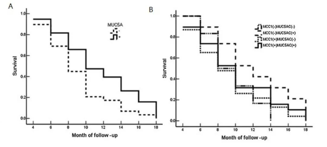
Figure 5. MUC5AC expression and survival curves in gallbladder adenocarcinoma patients (A: Analysis of MUC5AC expression and overall survival; B: Analysis of MUC5AC and MUC1 (also known as EMA) expression and overall survival)
In recent years, some scholars have also studied the expression of MUC5AC in tumors such as esophageal cancer, parotid gland cancer, nasopharyngeal cancer, and pancreatic cancer. They believe that negative expression of MUC5AC is a risk factor affecting the prognosis of esophageal cancer patients; positive expression of MUC5AC may be associated with a good prognosis in parotid gland cancer and a poor prognosis in nasopharyngeal cancer; the relationship between MUC5AC expression in pancreatic cancer and prognosis is still controversial and requires further in-depth research.
In summary, MUC5AC has different abnormal expressions in gastric cancer, colorectal cancer, lung cancer, and gallbladder lesions, and is closely related to tumor prognosis:
-
Downregulation of MUC5AC expression in gastric cancer is involved in its occurrence and development. It is an independent factor for predicting gastric cancer prognosis. High expression of MUC5AC indicates better differentiation of gastric cancer and possibly a better prognosis.
-
MUC5AC expression in colorectal cancer is a potential marker for good prognosis and a therapeutic target.
-
MUC5AC expression in non-small cell lung cancer plays an important role in invasion and metastasis. Positive expression of MUC5AC is an independent factor for poor prognosis and may be a potential therapeutic target.
-
Downregulation of MUC5AC expression in gallbladder lesions is closely related to the pathogenesis of gallbladder tumors and is an independent prognostic predictor for gallbladder adenocarcinoma: downregulation of expression is positively correlated with decreased overall survival, indicating a poorer prognosis.
Related Antibodies from Maixin
|
Antibody Name
|
Product Number
|
Clone Number
|
Cellular Localization
|
|
MUC5AC
|
MAB-0079
|
45M1
|
Cytoplasm
|
References
[1]Yang J . Identification of novel biomarkers, MUC5AC, MUC1, KRT7, GAPDH, CD44 for gastric cancer[J]. Medical Oncology, 2020, 37(5).
[2] Zhang Yuefan, Zhang Weijie, Hu Jiajun, et al. Expression of MUC5AC and CDX-2 in Gastric Cancer Tissues and Their Correlation with Clinicopathological Features and Prognosis, 2020, 21(19):2282-2285.
[3]Hazgui M , Weslati M , Boughriba R , et al. MUC1 and MUC5AC implication in Tunisian colorectal cancer patients[J]. Turkish Journal of Medical Sciences, 2021 , 51: 309-318.
[4]Mesa H , Manivel J C , Larson W S , et al. Immunophenotypic Comparison of Neoplasms of the Appendix, Right Colon, and Left Colon in Search of a Site-Specific Phenotypic Signature[J]. International journal of surgical pathology, 2019,28(1): 20-30.
[5] Dong Y , Zhou L , Zhao D , et al. MUC5AC enhances tumor heterogeneity in lung adenocarcinoma with mucin production and is associated with poor prognosis[J]. Japanese Journal of Clinical Oncology, 2020,50(6):701–711.
[6] Wu Qiang, Zhang Xianghai. Clinical Significance of CD147 and MUC5ac Expression in Peripheral Non-Small Cell Lung Cancer and Their Correlation with CT Signs [C]// National Symposium on Clinical Experience in Integrated Traditional Chinese and Western Medicine for Liver Disease Treatment. World Federation of Chinese Medicine Societies; Journal of Traditional Chinese Medicine Press, 2017, 19(4):549-551.
[7]A B hoge. Immunohistochemical Study of MUC1 and MUC5AC Expression in Gall Bladder Lesions[J]. Journal of Clinical & Diagnostic Research, 2017, 11(7):12-16.
[8]Li X , Yang Z , Yang L , et al. Expressive levels of MUC1 and MUC5AC and their clinicopathologic significances in the benign and malignant lesions of gallbladder.[J]. Journal of Surgical Oncology,
[9] Song Zhenguo, Zhang Zhandong, Su Yonglin, et al. Correlation between MUC1 and MUC5AC Expression Levels and Prognosis of Esophageal Cancer [J]. Journal of Tropical Medicine, 2018, 18(7):902-906.
[10] Jiang Weidong, Li Wenhui, Yu Liqiao, et al. Expression and Clinical Significance of MUC1 and MUC5AC in Parotid Gland Cancer [J]. Journal of Xuzhou Medical University, 2019, 39(3):6166-171.
For more information, please contact: 800-8581156 or 400-889-9853
