
Maimai’s Pathology Weekly Reading Notes Issue 8 | Immunohistochemical Markers for Respiratory and Lung Tumors (Part 2)
Preface:
Based on the content arrangement of the book “Immunohistochemistry in Tumor Diagnostics” published by Springer, this article “Mai Mai” will focus on compiling and introducing knowledge points related to immunohistochemical markers for thymic epithelial tumors, as well as cardiac and pericardial tumors.tocheTEXT_3

Immunohistochemical Markers for Respiratory Tract and Lung Tumors
1. Immunohistochemical markers for thymic epithelium, such as Pax-8, CD117, CD5, CK (high molecular weight), p63.
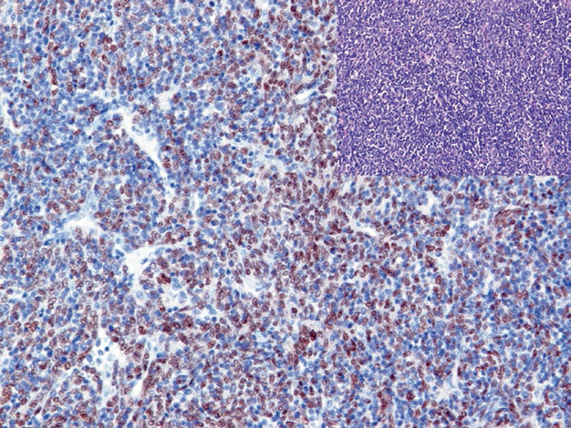
Figure 1. Type AB thymoma, Pax-8 shows positive nuclear expression in neoplastic epithelial cells.

Figure 2. Thymic carcinoma, Pax-8 shows positive nuclear expression in malignant thymic epithelial cells.

Figure 3. Thymic carcinoma, CD117 shows positive expression in malignant thymic epithelium.

Figure 4. In type AB thymoma, CK5/14 shows positive cytoplasmic expression in neoplastic epithelial cells.
2. Immunohistochemical markers for thymic lymphoid stroma, such as CD1a, CD3, TdT.
This part will be detailed in the section on lymphohematopoietic-related tumors, and refer to Table 1.
Table 1. Summary of Immunohistochemical Indicators for Thymic Epithelial Tumors

Notes for Table 1:
-
Type A and AB thymomas may also show positive expression of CD20;
-
In spindle cell thymic carcinoma, CD5 is negative.
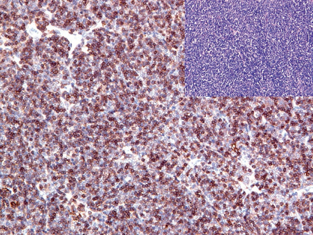
Figure 5. In type AB thymoma, tumor-associated T lymphocytes are positive for CD1a.
Immunohistochemical Markers for Cardiac and Pericardial Tumors
Table 2. Summary of Immunohistochemical Indicators for Cardiac and Pericardial Tumors
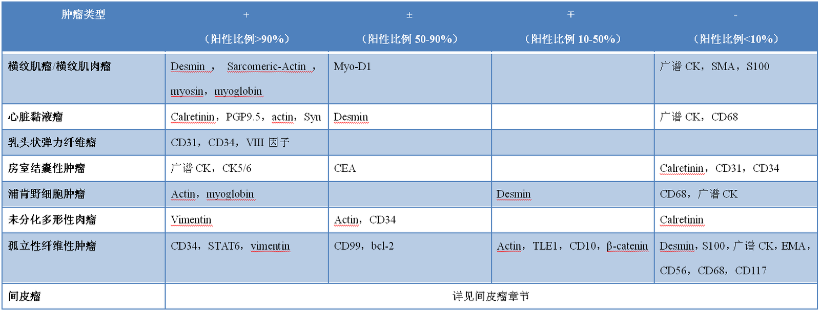

Figure 6. Cardiac papillary fibroelastoma, vascular endothelial cells show positive immunohistochemical staining for CD31.
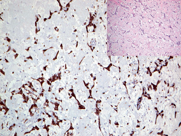
Figure 7. Cardiac myxoma, tumor cells strongly express calretinin.
1
Mai Mai: “Technology empowerment is the main theme of the era. Mastering core technologies and not being constrained by others is a prerequisite for innovative enterprises. In the fourth issue of “Mai Mai Reading,” we introduced the Maxin CloneDomesticCKseries of antibodies, which have received attention and praise from many pathology teachers. Today, we recommend several Maxin Clone antibodies related to the content of this article. You might want to try them when you have the opportunity.”
|
Antibody Name |
Clone Number |
Positive Control |
Positive Location |
|
Actin |
MX083 |
Colonic smooth muscle, appendix |
Cytoplasm |
|
Bcl-2 |
MX022 |
Tonsil, appendix |
Membrane/cytoplasm |
|
CD5 |
MX052 |
Tonsil, thymus |
Membrane |
|
CD10 |
MX002 |
Tonsil, kidney | Cytoplasm/membrane |
|
CD31 |
MX032 |
Hemangioma, liver |
Membrane |
| CD56 | MX039 | Small cell lung carcinoma, appendix |
Membrane |
|
CD68 |
MX075 |
Malignant fibrous histiocytoma, tonsil |
Cytoplasm |
| CEA | MX068 | Lung adenocarcinoma, appendix |
Cytoplasm |
|
CgA |
MX018 |
Pheochromocytoma, neuroendocrine carcinoma |
Cytoplasm |
| Desmin | MX046 | Esophageal tissue, appendix |
Cytoplasm |
|
HER-2 |
MXR001 |
Invasive ductal carcinoma of the breast |
Membrane |
| MyoD1 | MX049 |
Rhabdomyosarcoma |
Nucleus |
|
p63 |
MX013 |
Prostatic hyperplasia, urothelial carcinoma |
Nucleus |
| Pax-8 | MX062 |
Renal clear cell carcinoma, fallopian tube | Nucleus |
|
SMA |
MX097 |
Colonic smooth muscle, appendix |
Cytoplasm |
| Syn | MX038 | Pancreas, appendix | Cytoplasm |
|
Vimentin |
MX034 |
Gastrointestinal stromal tumor, appendix |
Cytoplasm |
For more information, please contact: 800-8581156 or 400-889-9853