
Maimai’s Pathology Weekly Reading Notes Issue 16 | Immunohistochemical Markers for Diagnosing Female Genital Tract Tumors (Part 2)
Preface:
In the previous article, we mainly introducedFemale reproductive tract organsthe commonly used immunohistochemical markers for tumors of the external genitalia and vagina, cervix and uterine body, as well as fallopian tubes and uterine ligaments. In this issue, we will systematically review and explain theCommonly used immunohistochemical markersof various ovarian tumors, hoping that through such introductions, we can provide some reference for pathology colleagues in selecting corresponding tumor immunohistochemical markers.

Table 1. Commonly used immunohistochemical markers for ovarian epithelial tumors
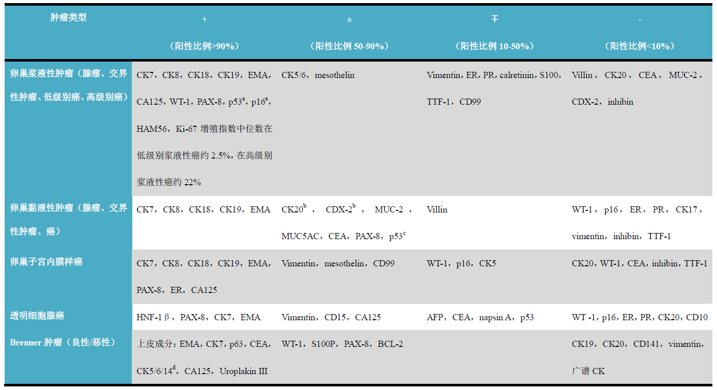
(Click to view larger image)
Note:
-
High expression is characteristic in high-grade serous carcinoma, while low expression or negative in low-grade carcinoma;
-
CDX-2 and CK20 are positive in mucinous adenocarcinoma and intestinal-type adenoma;
-
Generally negative in adenoma and borderline tumors;
-
Basal epithelial cells express CK5/6/14.
Table 2. Commonly used immunohistochemical markers for ovarian sex cord-stromal tumors
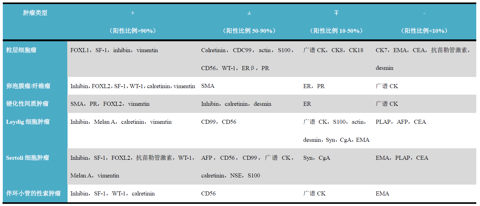
(Click to view larger image)
Table 3. Commonly used immunohistochemical markers for ovarian germ cell tumors
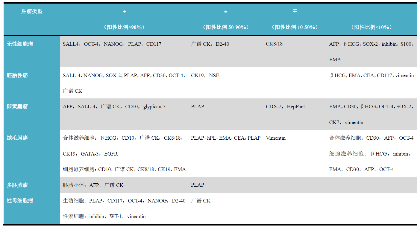
(Click to view larger image)
Table 4. Commonly used immunohistochemical markers for other ovarian tumors
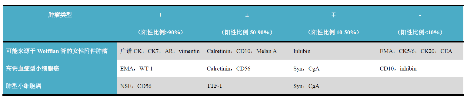
(Click to view larger image)
Note:
-
In mucinous endometrioid adenocarcinoma,CDX-2can be positive;
-
p16andp53positivity is generally only seen in leiomyosarcoma;
-
In placental site nodule and exaggerated placental site,Ki-67proliferation index<1%, in choriocarcinomaKi-67proliferation index>50%。
Detailed explanation of some markers
WT-1
Positive expression pattern: nuclear
Recommended positive control tissue: Wilms tumor, mesothelioma, etc.
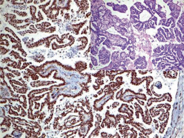
Figure 1. Ovarian serous carcinoma, immunohistochemistry shows strong nuclear positivity for WT-1.
CA125
Positive expression pattern: cell membrane (luminal)
Recommended positive control tissue: ovarian serous carcinoma
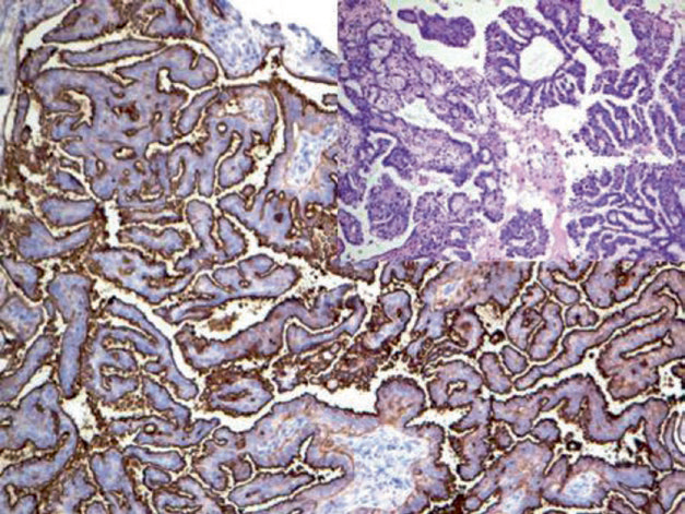
Figure 2. Ovarian serous carcinoma, immunohistochemistry shows cell membrane staining for CA125.
FOXL2
Pax-8
1
Maimai: “CA 125 is a major marker for epithelial ovarian cancer, especially serous carcinoma, and has reference value in the differential diagnosis of serous carcinoma and mucinous carcinoma. Pax-8 is highly expressed in ovarian serous carcinoma but almost not expressed in ovarian mucinous carcinoma. Therefore, the positive expression of both has its specific reference significance. Besides the important roles mentioned above, the WT-1 marker can also be used in the differential diagnosis of some small round cell tumors, etc.””
|
Antibody Name |
Clone Number |
Positive Control |
Positive Location |
|
CA 125* |
MX055 |
Ovarian serous carcinoma, fallopian tube |
Cytoplasm/Cell membrane |
|
Pax-8* |
MX062 |
Renal clear cell carcinoma, fallopian tube |
Nucleus |
|
WT-1* |
MX012 |
Kidney tissue, Wilms tumor |
Nucleus |
*Marked as Maxim clone products
For more information, please contact: 800-8581156 or 400-889-9853