
IHC Detection of MMR Protein Series Tweets – Interpretation of MMR Protein Detection Results
Introduction:
In the previous issue, it was mentioned that due to factors such as different detection methods, dMMR and MSI detection results are not completely equivalent. IHC detection of MMR proteins has advantages such as strong accessibility, simplicity, and the ability to indicate mutated genes, making it an irreplaceable method for LS screening. However, the IHC method also has certain limitations. How to interpret the staining results of the four antibodies? How to interpret the expression patterns of MMR proteins? How to resolve misinterpretations of results? This issue provides a brief overview of the above questions.
I. Staining and Interpretation of MMR Proteins in Colorectal Cancer
-
Under normal conditions, the four MMR proteins (MLH1, MSH2, MSH6, PMS2) stain in the nuclei of colorectal cancer cells, while the nuclei of internal controls (lymphocytes and normal intestinal mucosal epithelial cells within the tissue) also show staining. If patchy nuclear staining, cytoplasmic staining, nuclear membrane staining, or nucleolar staining occurs, further examination is recommended.
-
Any nuclear expression in tumor cells is determined as positive for that MMR protein. Typically, the nuclear staining intensity in most colorectal cancer cells is equivalent to (or even higher than) that of the internal control cells, and interpretation can be combined with internal and external control results.
-
The American Society of Pathology recommends using the classification methods “loss of expression” and “normal expression” for MMR detection reports, rather than “negative” and “positive,” as the latter can easily lead to misunderstandings. Many may think “negative” is normal, whereas in fact, a “negative” result may indicate LS. For cases where IHC is inconclusive, it is recommended to report as “further examination advised.”
II. Interpretation of MMR Protein Expression Patterns in Colorectal Cancer

Note: Further testing helps in differentiation, excluding Lynch-like syndrome, and conducting monitoring and follow-up for family members.
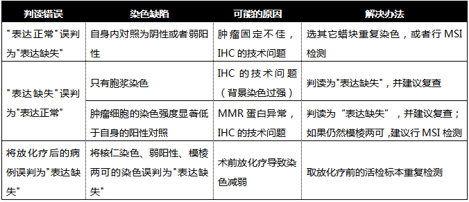
III. Causes and Solutions for Result Misinterpretation

References:
[1] Wang HL, Kim CJ, Koo J, et al. Practical Immunohisto-chemistry in Neoplastic Pathology of the Gastrointestinal Tract, Liver, Biliary Tract, and Pancreas[J].Archives of pathology & laboratory medicine,2017,141(9):1155-1180
[2] Gao Xianhua, Zhang Wei, Bai Chenguang. Deficiencies and coping strategies of immunohistochemistry in screening Lynch syndrome [J/CD]. Chinese Journal of Colorectal Diseases (Electronic Edition), 2019, 8(5): 439-446. DOI: 10.3877/cma.j.issn.2095-3224.2019.05.002