
Fifteenth Issue of “Maimai” Pathology Weekly Reading Notes | Immunohistochemical Markers for Diagnosing Female Reproductive Tract Tumors (Part 1)
Preface:
Malignant tumors can occur in almost all parts of the female reproductive tract, with cervical cancer being the most common, followed by others such as endometrial cancer and malignant ovarian tumors. With the deepening of research on traditional markers and the continuous discovery of new markers, immunohistochemistry plays an increasingly important role in the pathological diagnosis of female reproductive tract tumors. We have briefly summarized this content, which will be introduced in two parts, hoping to assist pathology colleagues in their diagnostic work.

Table 1. Commonly Used Immunohistochemical Markers for Vulvar and Vaginal Tumors

(Click to view larger image)
Table2. Commonly Used Immunohistochemical Markers for Cervical Tumors
(Click to view larger image)
Note:
-
Pax-2Generally expressed in benign hyperplastic endocervical glands;
-
In glandular componentsCK7positive;
-
In squamous componentsCK5/6、CK14positive.
Table 3. Commonly Used Immunohistochemical Markers for Uterine Body Tumors

(Click to view larger image)
Note:
-
In mucinous endometrial adenocarcinoma,CDX-2can be positive;
-
p16andp53positivity is generally only seen in leiomyosarcoma;
-
In placental site nodule and placental site overreaction,Ki-67proliferation index<1%, in choriocarcinomaKi-67proliferation index>50%。
Table 4. Commonly Used Immunohistochemical Markers for Fallopian Tube and Uterine Ligament Tumors

Note:
-
Diffusely expressed in serous tubal intraepithelial carcinoma, but only a few scattered cells express it in normal fallopian tube mucosa.
Detailed Explanation of Some Markers
p16
Positive expression pattern: Nuclear/Cytoplasmic
Recommended positive control tissue: Cervical squamous cell carcinoma
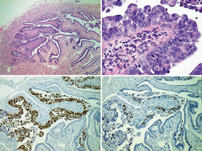
Figure 1. Serous tubal intraepithelial carcinoma, HE-stained section shows significant atypia in the fallopian tube epithelium; on consecutive section immunohistochemical detection, these cellsp53show strong nuclear positivity, Ki-67proliferation index about 15%。
Pax-8
HNF1β
PTEN
Steroid hormone receptors
1
Mai Mai: “p16 is closely related to HPV infection-induced cervical cancer and can also assist in the differential diagnosis of serous carcinoma from other types of tumors. In the diagnosis of high-grade lesions, p16 expression is similar to histological diagnostic criteria, improving the accuracy of pathologists’ diagnoses and more accurately predicting the risk of high-grade lesions. Current research finds that Pax-8 is expressed in some subtypes of kidney, bladder, and thyroid tumors and in ovarian malignancies, but not in most epithelial tumors such as breast cancer, lung cancer, and malignant mesothelioma. Therefore, Pax-8 can serve as an effective marker for determining the primary site of tumors and for tumor differential diagnosis. Additionally, compared to uterine serous carcinoma, high-grade endometrioid carcinoma commonly shows PTEN loss of expression, thus it can be used for differential diagnosis between uterine serous carcinoma and endometrioid carcinoma. HNF1β is a good marker for ovarian clear cell carcinoma and can also serve as a marker for testicular germ cell tumors; combined use with Glypican-3 can help differentiate yolk sac tumor from other germ cell tumors.””
|
Antibody Name |
Clone Number |
Positive Control |
Positive Location |
|
p16* |
MX007 |
Cervical squamous carcinoma, tonsil |
Nuclear/cytoplasmic |
|
Pax-8* |
MX062 |
Renal clear cell carcinoma, fallopian tube |
Nuclear |
|
PTEN |
6H2.1 |
Prostatic hyperplasia |
Nuclear |
|
HNF1β* |
MX092 |
Renal clear cell carcinoma, appendix |
Nuclear |
*Marked as Maxim clone product
For more information, please contact: 800-8581156 or 400-889-9853