Hotspot Focus

Application and Pitfalls of Immunohistochemical Antibodies in Diagnosing Urogenital Diseases (Part 2)


Introduction:


The application of immunohistochemical antibodies in the genitourinary system is becoming increasingly common. When biopsy tissue is very limited, selecting the optimal IHC markers becomes even more important. In the previous article, we shared the application and pitfalls of immunohistochemistry in diagnosing prostate diseases. This article will focus on the application and pitfalls of immunohistochemistry in diagnosing bladder diseases.


Bladder

Urothelial carcinoma (UC), as the most common histological type of bladder cancer, can exhibit various histological structures, such as glandular, squamous, or micropapillary features. Additionally, the bladder is the most common site for secondary malignant tumors (e.g., from the colon and prostate) to metastasize to the urinary system. Therefore, the differential diagnosis of bladder tumors also presents challenges, requiring immunohistochemical assistance. In addition to some prostate markers, commonly used markers for bladder tumors include GATA3, p63, CK20, etc. (Table 1).
These markers show positive expression in UC, although they share a common drawback: none are urothelial-specific, nor are they 100% sensitive. High-grade UC often shows patchy expression of CK20 or p63, or even complete loss of CK20 or p63. Subtypes of UC, including those with micropapillary components or glandular differentiation, may mimic the morphological appearance of tumors from other organs. These cases may also lose expression of CK20 and p63. Although staining may be weak and mottled, in most cases UC still retains expression of GATA3 (Figure 1, D). Second-line urothelial markers (such as Uroplakin III and thrombomodulin) have limited utility due to poor sensitivity for high-grade tumors.

TableBladder Markers

Marker

Benign Reactive Urothelium

Urothelial Carcinoma

Bladder Adenocarcinoma

CK20

Umbrella Cells

P63

GATA3

CD44

Full-thickness

/

p53

Basal Cells

Strongly Positive

/

β-Catenin

Non-nuclearstaining

Non-nuclear Staining

Non-nuclear Staining

CDX2


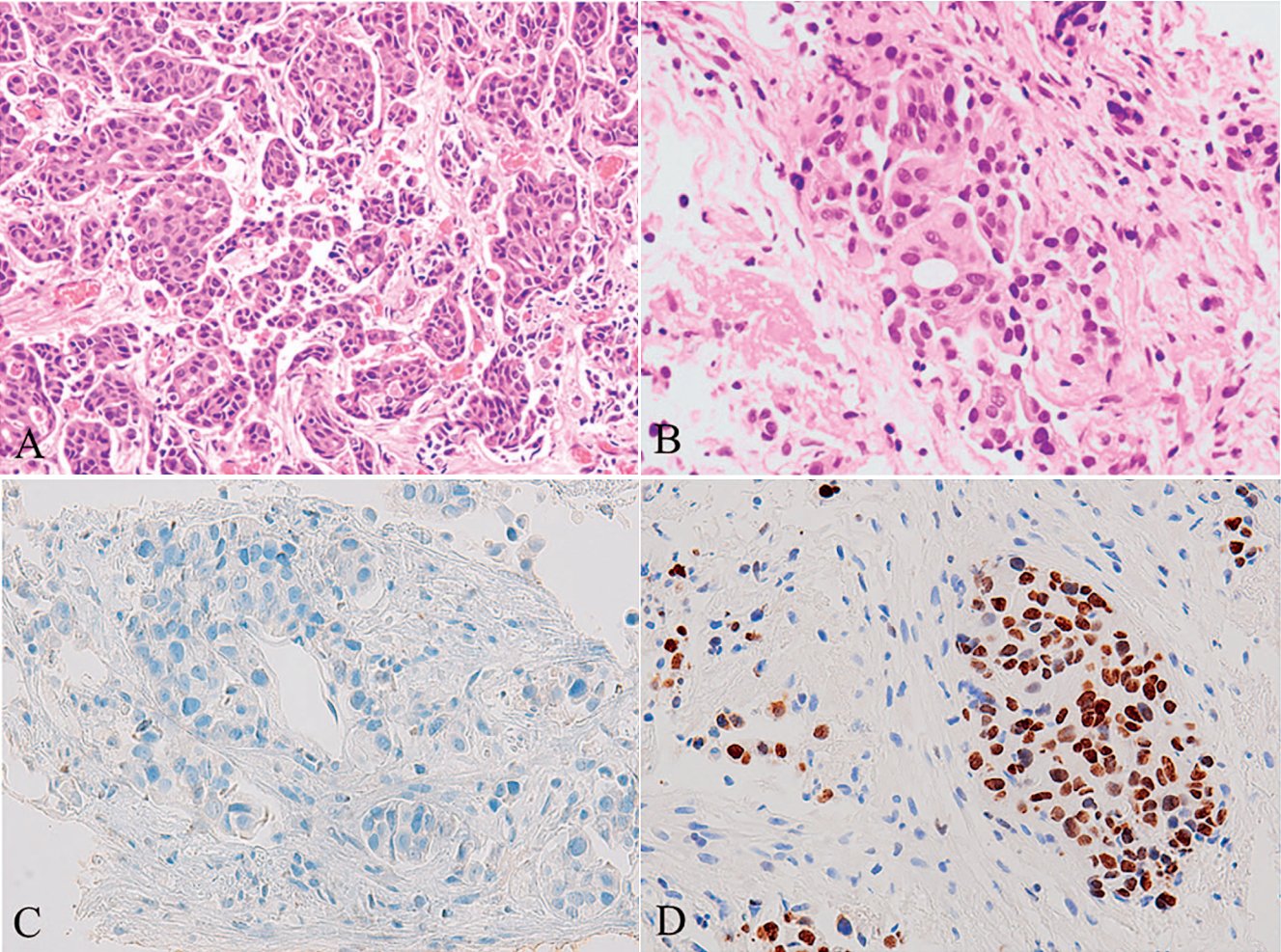
Figure 1A. Bladder Micropapillary Urothelial Carcinoma

Figure 1B. Bladder Micropapillary Urothelial Carcinoma. Clusters of tumor cells are embedded in ‘lacunae,’ and these tumor cells are relatively bland in morphology.

Figure 1C. Bladder Micropapillary Urothelial Carcinoma. p63 staining shows loss of basal cells.

Figure 1D. Bladder Micropapillary Urothelial Carcinoma. Positive expression of GATA3.


For the anatomical site of the bladder, secondary adenocarcinoma is far more common than primary bladder adenocarcinoma. This is especially true for metastases or direct spread from colorectal adenocarcinoma, prostate cancer in males, or female genital system tumors. Typically, prostate adenocarcinoma expresses prostate markers such as PSA and NKX3.1; female genital system tumors express PAX8, WT1, ER/PR, Vimentin, or p16. The immunoprofile of primary bladder adenocarcinoma (intestinal type) (Figure 2) is similar to that of colorectal adenocarcinoma: CDX2+, CK20+, and CK7-. In such cases, β-catenin can be used for differential diagnosis. Colorectal adenocarcinoma shows strong nuclear staining for β-catenin, whereas most primary bladder adenocarcinomas are negative for nuclear β-catenin (Figure 2, D). Cytoplasmic or membranous staining of β-catenin in bladder adenocarcinoma should not be interpreted as positive.

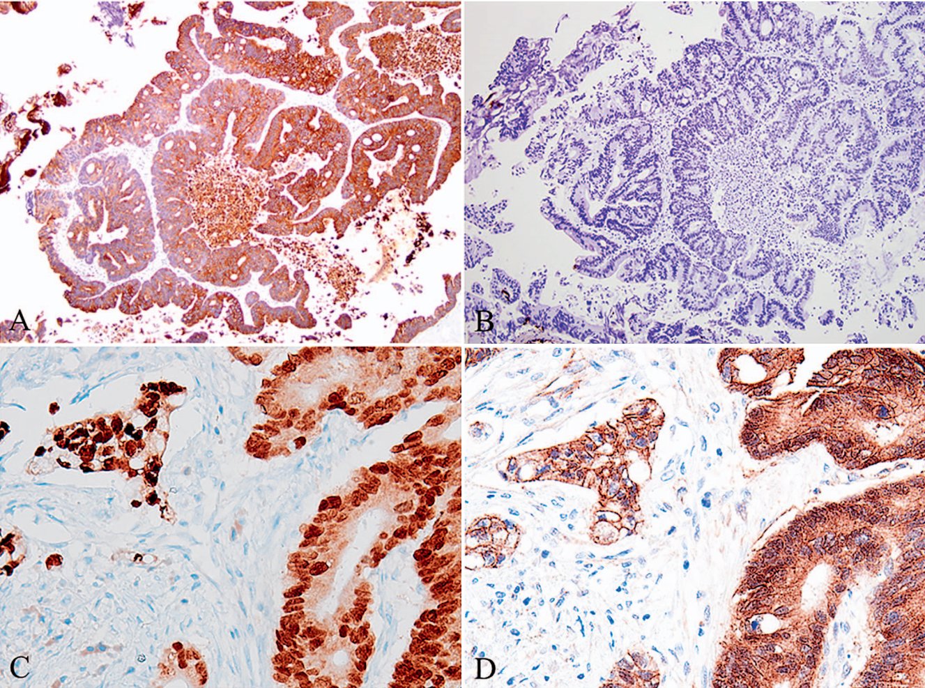
Figure 2A. Primary Bladder Adenocarcinoma. Similar to the immunophenotype of colorectal cancer, primary bladder adenocarcinoma (intestinal type) is CK20 positive;

Figure 2B. Primary Bladder Adenocarcinoma. Similar to the immunophenotype of colorectal cancer, primary bladder adenocarcinoma (intestinal type) is CK7 negative;

Figure 2C. Primary Bladder Adenocarcinoma. Similar to the immunophenotype of colorectal cancer, primary bladder adenocarcinoma (intestinal type) is CDX2 positive;

Figure 2D. Primary Bladder Adenocarcinoma. Primary bladder adenocarcinoma shows cytoplasmic staining for β-catenin, but lacks the nuclear β-catenin staining commonly seen in colorectal cancer.


In rare cases, certain benign bladder diseases can mimic malignancy. Viral infections may cause significant cytological atypia resembling malignancy (Figure 3, A and B). Typically, these cells may express strong p53 positivity and show high Ki-67 proliferative activity (Figure 3, C), mimicking carcinoma in situ UC. However, upon close observation, the chromatin has a uniform, glassy texture. Such cells are sometimes referred to as ‘decoy’ cells in cytology but are rarely seen in biopsy specimens. In specimens infected with BK virus, immunohistochemistry using a specific antibody against SV40 (simian virus 40, a polyomavirus) can label the virus-infected cells.

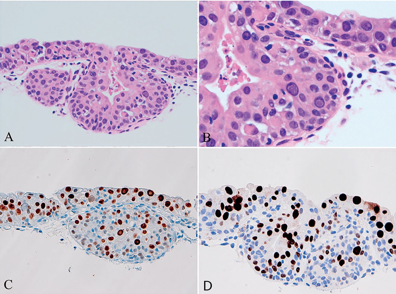
Figure 3A. Cytological Atypia due to BK Virus. Urothelial cells infected with BK virus;

Figure 3B. Cytological Atypia due to BK Virus. Marked cytological atypia, mimicking urothelial carcinoma in situ;

Figure 3C. Cytological Atypia due to BK Virus. Very high proliferative activity of Ki-67;

Figure 3D. Cytological Atypia due to BK Virus. Positive staining with SV40 antibody cross-reactive with BK virus.


For biopsy or transurethral resection specimens, assessing the presence of the muscularis propria and/or tumor invasion into the muscularis propria is crucial for clinical staging. For specimens with severe physical damage, determining the presence of smooth muscle tissue can be quite difficult. Among all smooth muscle markers, smoothelin performs exceptionally well. Smoothelin is a novel cytoskeletal smooth muscle-specific protein, expressed almost exclusively in terminally differentiated (contractile) smooth muscle cells. Smoothelin can show different staining intensities: smoothelin staining is stronger in the muscularis propria compared to the muscularis mucosae or arterial walls. When neither type of smooth muscle is present in the biopsy specimen or there is physical damage such as cautery, identification becomes difficult. In such cases, small arterial smooth muscle can be used as a reference for judging smoothelin expression intensity. Smoothelin expression stronger than that in background small arteries usually indicates muscularis propria, while weaker staining indicates muscularis mucosae (Figure 4, A). This can assist pathologists in determining whether UC invades the muscularis propria, which is often an indication for radical cystectomy.

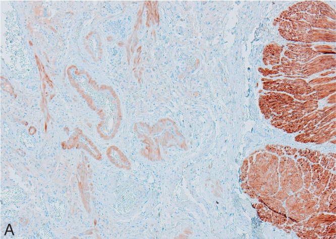
Figure 4A. Application of Smoothelin in Bladder Cancer. Smoothelin is strongly positive in the muscularis propria (right side of Figure 8A, Smoothelin stain) and weakly positive in the muscularis mucosae and smooth muscle bundles of background vessels (left side of Figure 8A, Smoothelin stain).

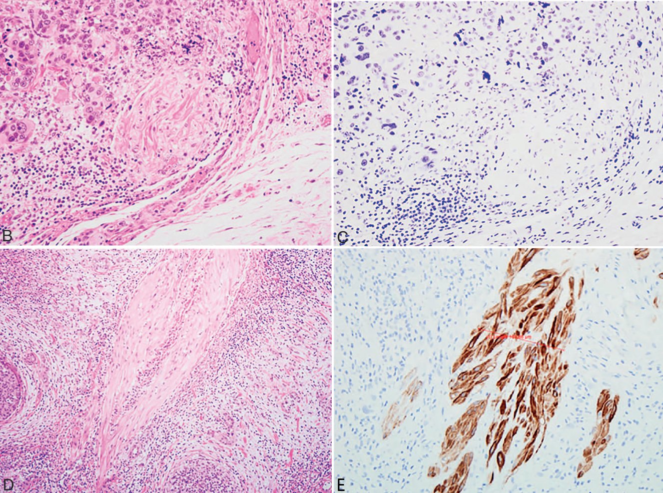
Figure 4B. Application of Smoothelin in Bladder Cancer. Urothelial carcinoma cells invading smooth muscle bundles;

Figure 4C. Application of Smoothelin in Bladder Cancer. Same field as Figure 8B, showing faint smoothelin staining, suggesting involvement of the muscularis mucosae;

Figure 4D. Application of Smoothelin in Bladder Cancer. Another case of urothelial carcinoma invading smooth muscle bundles;

Figure 4E. Application of Smoothelin in Bladder Cancer. Same field as Figure 8D, showing strong smoothelin positivity, suggesting involvement of the muscularis propria.


Maxin Related Antibodies


Antibody Name

Product Code

Clone Number

Positive Localization

CK20*

MAB-0834

MX059

Cytoplasmic

CDX2*

MAB-0713

MX024

Nuclear

p53*

MAB-0674

MX008

Nuclear

p63*

MAB-0694

MX013

Nuclear

GATA3

MAB-0695

L50-823

Nuclear

β-Catenin*

MAB-0754

MX043

Cytoplasmic/Membranous

*Indicates Maxin clone product


References:

Jenny Ross, Guangyuan Li, Ximing J. Yang. Application and Pitfalls of Immunohistochemistry in Diagnosis of Challenging Genitourinary Cases. Arch Pathol Lab Med. Vol 144, March 2020:290-304

发表回复

您的邮箱地址不会被公开。 必填项已用 * 标注