Common Immunohistochemical Markers for Skin Tumors
Common immunohistochemical markers for epidermal tumors include certain CKs, EMA, Ep-CAM, p16, p53, HPV, and Ki-67; for sweat gland tumors, common markers include certain CKs, p63, CEA, EMA, CD15, GATA3, S100, ER, PR, AR, and GCDFP-15; for hair follicle tumors, common markers include certain CKs, p63, EMA, hair keratin (HKN), human hair keratin (HHK), and Ep-CAM; for sebaceous gland tumors, common markers include certain CKs, EMA, Ep-CAM, AR, adipophilin, and perilipin.
Table 1. Overview of Common Immunohistochemical Markers for Epidermal Tumors

(Click to view larger image)
Table 2. Overview of Common Immunohistochemical Markers for Skin Appendage Tumors

(Click to view larger image)
Note:
-
EMA positivity is more commonly seen in malignant tumors.
Table 3. Overview of Common Immunohistochemical Markers for Other Skin Tumors
(Click to view larger image)
Note:
-
EMA positivity is more commonly seen in malignant tumors;
-
Perinuclear comma-shaped staining;
-
Nuclear staining.
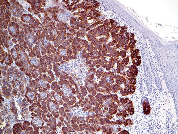
Figure 1. Basal cell carcinoma, strongly positive for Ep-CAM. Note that epidermal cells in the image do not express this marker.
Similar to normal sweat glands, the tumor cells of eccrine and apocrine sweat glands are composed of the same cell types; generally, they consist of luminal cells and basal/myoepthelial-type cells, although the distribution and morphology of these two cell types vary to some extent in tumors of different grades. Because there are two cell types, the immunohistochemical characteristics of this group of tumors must consider both cell types. They generally express CEA and hormone receptors to some degree, commonly show GATA-3 positivity, and the intensity of positivity may differ between cell types. Additionally, many sweat gland tumors exhibit morphological and immunohistochemical features similar to salivary gland tumors, such as resembling adenoid cystic carcinoma.
Hair-specific keratins include hair keratins 5, 6, 7, and human hair keratin, which are specific for hair follicle tumors. Among various CKs, CK15 is relatively specific for hair follicles, nails, and hair follicle tumors. Meanwhile, CK15 is a marker for epidermal stem cells, and its expression in stratified squamous epithelium is limited to the basal layer. Sebaceous gland tumors generally do not express CK15.
As mentioned earlier, the immunohistochemistry of melanocytic tumors will be covered in a dedicated chapter. Merkel cell carcinoma commonly expresses immunohistochemical markers such as Merkel cell polyomavirus, EMA, CD56, and NSE.
Detailed Explanation of Some Markers
Positive expression pattern: Cell membrane/cytoplasm
Recommended positive control tissue: Skin
Adipophilin is a lipid droplet-associated protein expressed on the surface of lipid droplets in the cytoplasm of various types of normal cells, such as alveolar cells of lactating mammary glands, the zona fasciculata of the adrenal gland, and Sertoli cells, while adipocytes do not express this marker.
Tumor cells containing lipid droplets also express this marker, and it is specific for sebaceous gland tumors. Studies on other tumor types with clear cell morphology that resemble sebaceous gland tumors found that adipophilin positivity was 92% in sebaceous carcinoma, 100% in the studied sebaceous adenomas and xanthelasmas, and 65% in metastatic renal cell carcinoma; other tumors with clear cell features were all negative, such as squamous cell carcinoma, basal cell carcinoma, trichilemmoma, and clear cell hidradenoma.
Adipophilin can also be used to mark Burkitt lymphoma because its cytoplasm also contains lipid droplets.
Perilipin is another marker for sebaceous gland tumors and is also a lipid droplet-associated protein. Normally, it is expressed in adrenal cortical cells, Leydig cells, brown adipose tissue, and white adipose tissue cells; this marker localizes to the surface of lipid droplets and is involved in lipid metabolism.
Approximately one-third of sebaceous gland tumors express Perilipin, but it is not specific, as this marker can also be expressed in other tumor types with clear cell morphology.
MaiMai Recommends: “Skin tumors are proliferative diseases occurring in skin cells, clinically classified as benign, malignant, and precancerous dermatoses that are prone to malignant transformation. Immunohistochemistry aids in the diagnosis and differential diagnosis of skin tumors and is helpful for understanding andevaluating cutaneous lymphocytic infiltration.! This issue, MaiMai recommends the following antibodies, you might want to give them a try.”
|
Antibody Name
|
Clone Number
|
Positive Control
|
Cellular Localization
|
|
CEA*
|
MX068
|
Lung adenocarcinoma, appendix
|
Cytoplasm
|
|
EMA
|
E29
|
Esophageal squamous cell carcinoma, meningioma
|
Cytoplasm/cellmembrane
|
|
Ep-CAM*
|
MX066
|
Lung adenocarcinoma, kidney
|
Cell membrane
|
|
GATA3
|
L50-823
|
Urothelial carcinoma, breast cancer
|
Nucleus
|
*Marked as Maxin clone products
For more information, please contact: 800-8581156 or 400-889-9853