Overview of Immunohistochemistry for Vascular Tumors
Commonly used immunohistochemical markers for vascular tumor diagnosis include CD31, CD34, Factor VIII, CD105, ERG, podoplanin (D2-40), thrombomodulin (CD141), and Fli-1.
Table 1. Common Immunohistochemical Markers for Vascular Tumors

(Click to view larger image)
Note:
-
Glut-1 is generally positive in hemangiomas, but negative in vascular malformations, pyogenic granulomas, and granulation tissue;
-
Dabska tumor, also known as malignant endovascular papillary angioendothelioma or lymphangiomatous papillary angioendothelioma.
Detailed Explanation of Some Markers
Positive expression pattern: Cell membrane/cytoplasm
Recommended positive control tissue: Appendix
CD31, also known as platelet endothelial cell adhesion molecule-1 (PECAM-1), is a transmembrane glycoprotein belonging to the immunoglobulin family. It is normally expressed at endothelial cell junctions and on the surfaces of platelets, monocytes, granulocytes, and B lymphocytes.
CD31 is a sensitive and specific marker for blood vessels and vascular tumors. However, in rare cases, non-vascular tumors may also show low-level expression of CD31, such as certain lymphomas, plasmacytomas, Langerhans cell tumors, leiomyosarcomas, mesotheliomas, gliomas; and even a few carcinomas, such as in situ and invasive breast cancer, and papillary thyroid carcinoma.
Positive expression pattern: Cell membrane
Recommended positive control tissue: Appendix
CD34 is a cell surface adhesion glycoprotein expressed on precursor hematopoietic cells of the myeloid and lymphoid lineages, some mesenchymal stem cells, and endothelial cells, and is expressed in many tumors derived from these cells. Therefore, CD34 is one of the important markers for hematopoietic stem cells and mesenchymal stem cells, and can also mark myeloblasts in acute myeloid leukemia.
In clinical practice, CD34 is widely used as a marker for blood vessels and vascular tumors, but its specificity is not as high as CD31. CD34 is also an important marker for other tumors, such as dermatofibrosarcoma protuberans and GIST. However, due to its broad expression spectrum, it must be used in combination with other more specific markers.
Positive expression pattern: Cytoplasm
Recommended positive control tissue: Appendix
Factor VIII is a glycoprotein complex composed of three subunits, with functional domains corresponding to binding platelet glycoprotein, collagen, and heparin, respectively. Factor VIII is synthesized by vascular endothelial cells and megakaryocytes and stored in Weibel-Palade bodies of endothelial cells.
Factor VIII is a specific marker for blood vessels and vascular tumors, and its expression level is related to the differentiation grade of vascular tumors, with very low expression in poorly differentiated vascular tumors (such as angiosarcoma).
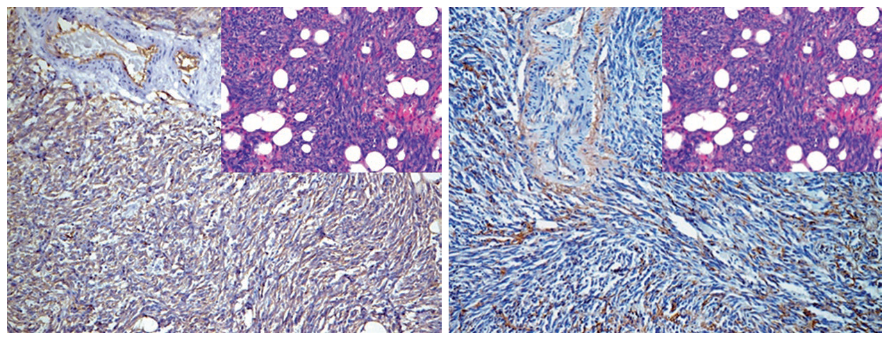
Figure 1. In angiosarcoma, CD34 (left) shows diffuse positivity, while Factor VIII (right) shows positive expression in most tumor cells.
Positive expression pattern: Cell membrane/cytoplasm
Recommended positive control tissue: Appendix
Podoplanin, also known as D2-40, is a type I transmembrane mucoprotein expressed on the cell membranes of fetal germ cells and certain mature cells, mainly lymphatic endothelial cells and mesothelial cells. In routine immunohistochemical testing, this marker is widely used to label lymphatic vessels, tumors of lymphatic endothelial origin, and mesotheliomas; in addition, it is an important marker for seminomas. However, its expression spectrum is relatively broad, and some tumors morphologically similar to the aforementioned tumors may also express this marker, such as leiomyosarcomas, desmoid tumors, peripheral nerve sheath tumors, etc. Therefore, in practice, it is necessary to add other more specific markers.

Figure 2. Endothelial cells in lymphatic vessels express D2-40, as seen in this case of lymphangitic carcinomatosis. However, note that Schwann cells in the nerve (top of the image) also show positive staining.
Positive expression pattern: Cell nucleus
Recommended positive control tissue: Blood vessel
ERG is one of the ETS family transcription factors, previously described in the prostate section. Normally, ERG is expressed in endothelial cells and is involved in regulating angiogenesis and endothelial cell apoptosis, making it an extremely sensitive and specific marker for endothelial cell tumors.
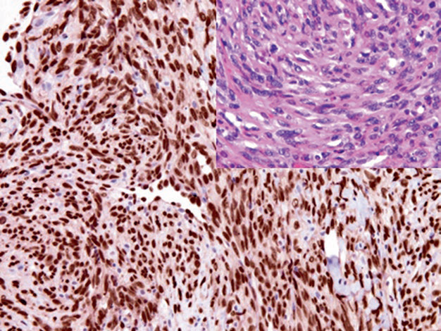
Figure 3. Angiosarcoma, with strong nuclear expression of ERG in tumor cells.
In addition to endothelial cell tumors, ERG is also expressed in some immature hematopoietic cells. In prostate cancer with TMPRSS2-ERG translocation, ERG is positive. For mesenchymal tumors, some other mesenchymal tumors morphologically similar to vascular tumors also express this marker, such as solitary fibrous tumors, fibrous meningiomas, and epithelioid sarcomas. In certain types of lymphomas, a small number of cases also show ERG expression.
HHV-8 is a DNA virus and is the causative agent of Kaposi sarcoma, primary effusion lymphoma, and multicentric Castleman disease. Detection of latent nuclear antigen is one of the diagnostic markers for Kaposi sarcoma.
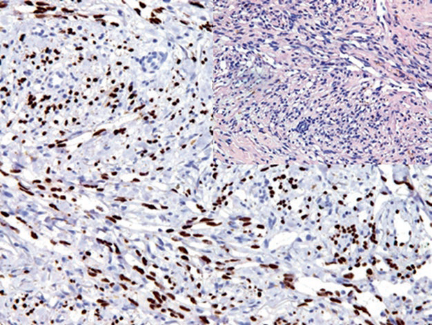
Figure 4. Expression of HHV-8 latent nuclear antigen in Kaposi sarcoma tumor cells.
MaiMai Recommendation: “Sometimes, we work hard to stain slides, but the staining results are not ideal. Why? Poor antibodies? This issue, MaiMai recommends the following antibodies to help you stain beautiful slides more easily.””
|
Antibody Name
|
Clone Number
|
Positive Control
|
Cellular Localization
|
|
CD31*
|
MX032
|
Hemangioma, Liver
|
Cell membrane
|
|
CD34
|
QBEnd/10
|
Hemangioma, Liver
|
Cell membrane/cytoplasm
|
|
D2-40
|
D2-40
|
Small intestine tissue, Mesothelioma
|
Cytoplasm/cell membrane
|
|
ERG*
|
MXR004
|
Angiosarcoma, Prostate cancer
|
Nucleus
|
|
HHV-8
|
13B10
|
Kaposi sarcoma
|
Cytoplasm/nucleus
|
*Marked as Maxin clone products
For more information, please contact: 800-8581156 or 400-889-9853