
Comparative Analysis of Specific Expression of Melanin-Related Antibodies
Melanoma-associated antigens (MAA’s) can be recognized by autologous T lymphocytes, including Melan-A/MART-1, MAGE-1, MAGE-3, Tyrosinase, gp100, gp75, BAGE-1, GAGE-1. The Melan-A/MART-1 antigen is expressed in over 90% of human melanomas and melanocytes. Except for retinal tissue, Melan-A/MART-1 RNA is not expressed in the vast majority of normal tissues, nor is it expressed in tumors arising from the colon, breast, brain, kidney, lung, bone, etc.
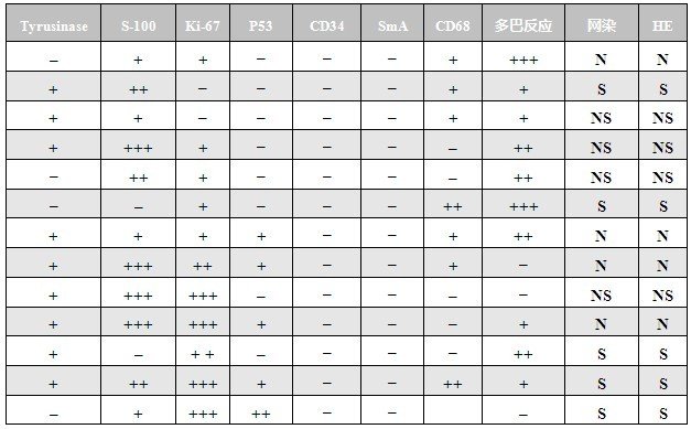
Expression of various melanoma markers and other markers in 12 cases of melanoma and 1 case of cutaneous metastatic hepatocellular carcinoma
Expression of various melanoma markers and other markers in 12 cases of melanoma and 1 case of cutaneous metastatic hepatocellular carcinoma
(This experimental result was provided by Su Qin)
Note: *, metastatic liver cancer; N, carcinoma-like area; S, sarcomatoid area; NS, area with both sarcomatoid and carcinoma-like features; immunohistochemical reaction intensity is judged as negative (-), weakly positive (-), moderately positive (++), and strongly positive (+++).
Note: *, metastatic liver cancer; N, carcinoma-like area; S, sarcomatoid area; NS, area with both sarcomatoid and carcinoma-like features; immunohistochemical reaction intensity is judged as negative (-), weakly positive (-), moderately positive (++), and strongly positive (+++).