Commonly used immunohistochemical markers for diagnosing endocrine and neuroendocrine tumors include CgA, Syn, NSE, S100, PGP9.5, CD56, Pax-6, synaptic vesicle protein 2, and somatostatin receptors.
It is important to note that the aforementioned immunohistochemical markers are commonly used to confirm neuroendocrine differentiation in normal or tumor tissues; however, none of these markers can be considered a universal marker for neuroendocrine differentiation. Therefore, in practical work, attention should be paid to the combined application of two or more antibodies.
Immunohistochemical markers commonly used for diagnosing neuroendocrine carcinomas are scattered across relevant chapters for different organs, such as small cell neuroendocrine carcinoma and large cell neuroendocrine carcinoma. Commonly used immunohistochemical markers include: various CKs, CgA, Syn, NSE, S100, CD56, somatostatin receptors, Ki-67; depending on the lesion site, site-specific markers such as CDX-2, SATB-2, PDX-1, Pax-6, TTF-1 can also be added.
Detailed Explanation of Some Markers
Positive Expression Pattern: Cytoplasm
Recommended Positive Control Tissue: Appendix
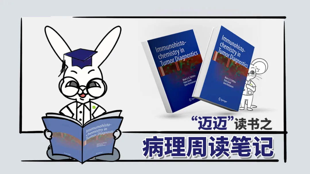
Chromogranin and Syn are the most commonly used immunohistochemical markers for neuroendocrine differentiation. Chromogranin is located within neurosecretory granules and synaptic vesicle walls in neuroendocrine cells, and includes chromogranin A (CgA), chromogranin B (also known as secretogranin I), and chromogranin C (also known as secretogranin II). CgA is the most commonly used marker in routine immunohistochemistry.
Almost all neuroendocrine cells and neuroendocrine tumors express chromogranin. The intensity of immunostaining depends on the amount of neuroendocrine granules in the cytoplasm of the examined cells. For example, although small cell carcinoma has strong synthetic activity for chromogranin, its cytoplasm is scant and neuroendocrine granules are sparse, so chromogranin immunostaining is generally very weak.
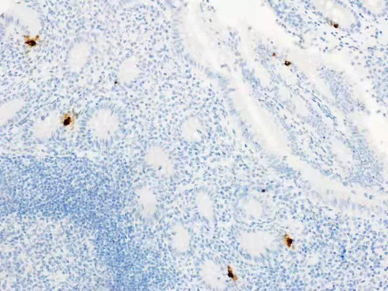
Figure 1. Appendix, CgA staining, positive in the cytoplasm of neuroendocrine cells.
Positive Expression Pattern: Cytoplasm
Recommended Positive Control Tissue: Appendix
Syn is a transmembrane calcium-binding glycoprotein and a major component of presynaptic vesicles. It is a broad-spectrum marker for neuroendocrine cells and tumors with neuroendocrine differentiation. Combined use of chromogranin and Syn enhances detection sensitivity.
Other synaptic vesicle proteins are rarely used in routine immunohistochemistry work, such as synaptic vesicle protein-2, synaptogranin, and vesicle-associated membrane protein.
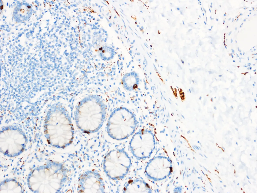
Figure 2. Appendix, Syn staining, positive in the cytoplasm of neuroendocrine cells, ganglion cells, and nerve plexus axons.
Positive Expression Pattern: Cytoplasm/Nucleus
Recommended Positive Control Tissue: Appendix
The full Chinese name of NSE is neuron-specific enolase. Enolase consists of α, β, and γ subunits forming homodimers or heterodimers. The antibody most commonly used in clinical practice targets the γ subunit. The γ subunit of NSE is mainly expressed in neurons, normal and neoplastic neuroendocrine cells, and can also be expressed to varying degrees in megakaryocytes, T cells, striated muscle cells, and smooth muscle cells.
However, it should be noted that NSE has low specificity for neuroendocrine tumors and is generally only used for screening; therefore, for the diagnosis of neuroendocrine tumors, support from other more specific markers is necessary.
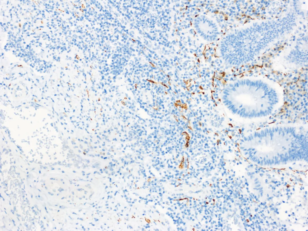
Figure 3. Appendix, NSE staining, positive in the cytoplasm of neuroendocrine cells and neuronal cells.
Positive Expression Pattern: Cytoplasm
Recommended Positive Control Tissue: Appendix
Normally, S100 is expressed in cells derived from the neural crest, such as glial cells, Schwann cells, melanocytes, chondrocytes, osteocytes, adipocytes, myoepithelial cells, dendritic cells, Langerhans cells, macrophages, and certain types of epithelial cells. In fact, the S100 protein family includes about 25 homologous low-molecular-weight cytoplasmic calcium-binding proteins, encoded by different genes located on different chromosomes. Precisely because of this, S100 is widely used, and the S100 antibody family has different monoclonal or polyclonal antibodies available for routine use.
In clinical practice, S100 is used as a screening marker with relatively poor specificity; therefore, for the diagnosis of neuroendocrine tumors, support from other more specific markers is necessary.
For other markers related to endocrine and neuroendocrine tumors, such as CD56, PGP9.5, etc., please refer to other chapters.
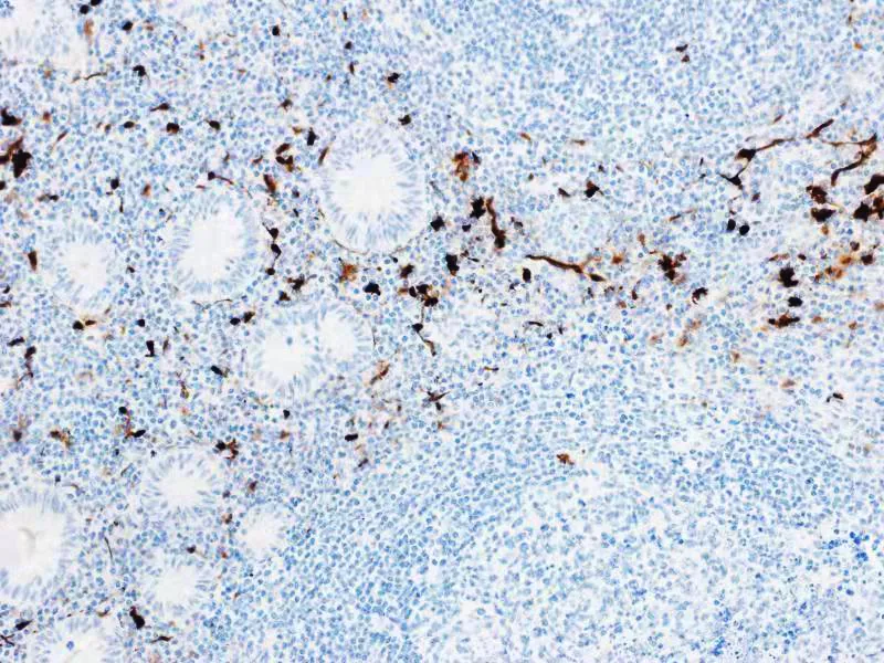
Figure 4. Appendix, S100 staining, positive in the nuclei and cytoplasm of Schwann cells of nerve fibers and ganglion satellite cells in the muscularis propria and submucosa of the appendix.1
MaiXin Recommendation:”A high-quality immunohistochemically stained slide provides a good foundation and prerequisite for correctly judging staining results, and the performance and quality of antibodies are key to high-quality immunohistochemically stained slides!””
|
Antibody Name
|
Clone Number
|
Positive Control
|
Cellular Localization
|
|
CgA *
|
MX018
|
Pheochromocytoma, Neuroendocrine Tumors
|
Cytoplasm
|
|
Syn*
|
MX038
|
Pancreas, Appendix
|
Cytoplasm
|
|
NSE
|
3-3-C
|
Pancreas, Appendix
|
Cytoplasm
|
|
S100
|
4C4.9
|
Schwannoma, Appendix
|
Cytoplasm/Nucleus
|
*Marked as MaiXin clone products
For more information, please contact: 800-8581156 or 400-889-9853