Hotspot Focus

A Conversation on Etiological Diagnosis


Preface:


Not long ago, due to the etiological diagnosis of a rare case, I intermittently discussed with several netizens skilled in microbiological examination on WeChat for a whole morning. This was a case of coccidioidomycosis, primarily caused by Coccidioides immitis infection. This disease is more common in the Americas, especially the southwestern United States; there are also a few sporadic cases in China. As of the end of 2018, only over 30 cases had been reported domestically, some of whom had traveled to areas where coccidioidomycosis is endemic.

This disease is mainly transmitted via the respiratory route, often occurring in populations with lower immune defense capabilities. It primarily affects the respiratory tract but can also disseminate, involving tissues such as the skin, central nervous system, kidneys, bones, and lymph nodes. The main lesions are suppurative and granulomatous. Etiological diagnosis can employ methods such as direct microscopic examination of specimens, histopathological examination, fungal culture, serological testing, and coccidioidin skin test. In terms of pathology, diagnosis is based on finding typical Coccidioides in the lesional tissue. Because it is quite rare, not only are many people in the microbiology department of the laboratory unfamiliar with it, but few pathologists know about it either. However, due to its distinctive morphology, once seen, it can be recognized. The author happened to encounter a case and, feeling it was special, consulted many Chinese and foreign literature, compared dozens of Chinese and foreign images, and finally confirmed Coccidioides. Therefore, I had some confidence and discussed it with several microbiology experts.


The conversation started with identifying several images (including tissue sections) they sent to WeChat, and we quickly reached a consensus on the pathogen. But someone asked,Can pathology confirm Coccidioides infection?Some agreed, some doubted. I said:If typical Coccidioides can be seen on pathological examination, a diagnosis can be made.Some friends in the laboratory field said,Infectious pathology is probably scarce nationwide.Pathology departments in large hospitals are more reliable.I cannot deny that within the pathology community, there are really not many physicians who value and are familiar with infectious pathology. The attention and identification ability of the pathology community towards pathogens are indeed weak. As they criticized, some pathology departments call any spore Cryptococcus and any hyphae Candida.Pathology departments generally do not use oil immersion lenses, so they often make mistakes. They even struggle with Helicobacter pylori, leading to their suspicion.As for Cryptococcus, Histoplasma, and Talaromyces marneffei (formerly called Penicillium marneffei) in lung tissue, doctors in general pathology departments cannot distinguish them.Therefore, they do not trust the Cryptococcus reported by pathology departments. There are also pathology departments suspecting Histoplasma, but the laboratory culture yields Talaromyces marneffei. For example, a certain tertiary hospital in Zhejiang cultured lung tissue puncture specimens; among over 2000 cases,many Cryptococcus were cultured (probably close to a hundred), and several cases of Talaromyces marneffei were also cultured, but not a single case of Histoplasma. Yet, the pathology department occasionally reports Histoplasma.They emphasized,For cases of fungal infection, there must be microbiological evidence, especially culture results.
The author believes,Indeed, histologically, these three (Cryptococcus, Histoplasma, Talaromyces marneffei) are indeed difficult to distinguish, so auxiliary technical examinations are needed, referencing microbiological tests, and gradually accumulating experience to become increasingly accurate.Diagnosing pathogens in infectious lesions,also depends on the experience and level of the diagnosing physician. I have always advocated that the diagnosis of infectious etiology should be combined with microbiological examination for greater certainty.Especially when experience is insufficient, it is particularly important to communicate with microbiology experts in the laboratory department.
Fortunately, some colleagues in the laboratory department (mainly referring to microbiological testing) have already noticed and are communicating with the pathology department, which is a promotion and help for us. They found that in their hospital,pathology department doctors only recognize Candida, Cryptococcus, Aspergillus, and Mucor as fungi.Moreover,The Mucor reported by pathology may not necessarily be correct; more likely it is Aspergillus. These two molds are sometimes difficult to distinguish. Theysometimes holding a skeptical attitude towards fungi reported in pathology reports is not necessarily a bad thing. Even common ones could be wrong. For example, Helicobacter pylori could be a false positive. They are particularly skeptical about the accuracy of some rare pathogens reported in pathology (reports).This skeptical attitude can remind us to carefully examine whether our diagnosis of pathogens is correct. I feel,given the current level of pathology departments, being able to distinguish these few types is already good; beyond that, they cannot differentiate.For example, Candida, Aspergillus, etc., still have many types. This is also a limitation of pathology, while fungal culture and genetic analysis can further classify. I advocate that pathologists and microbiologists should communicate more and learn from each other. This can avoid some low-level errors.The laboratory department has many subspecialties, but pathologists do not yet have a subspecialty in infectious pathology, and the pathology department does not have that many technical means and equipment.
During the conversation, I also learned that some laboratory colleagues are very interested in pathology; some also look at pathology slides;sometimes when they culture some rare fungi, they request pathology slides, mainly unstained sections, and stain them themselves. I think this is a good method.Some pathology departmentsdo not perform many special stains, often staining poorly and viewing poorly. Combining pathology and laboratory testing is very necessary.This way, they can complement each other and achieve mutual benefit.
In the United States, it is required to send infectious specimens simultaneously to pathology and microbiology for examination, cross-referencing each other, making etiological diagnosis much more reliable and accurate. Moreover, histopathology and microbiology belong to the same department of clinical pathology. In China, they are separate, so coordination is insufficient. According to the viewpoint of evidence-based medicine, seeking more evidence is necessary. The more evidence, the more reliable.Therefore, when performing etiological diagnosis, it is best to check whether the patient has corresponding microbiological, immunological, and molecular biological tests to corroborate each other.

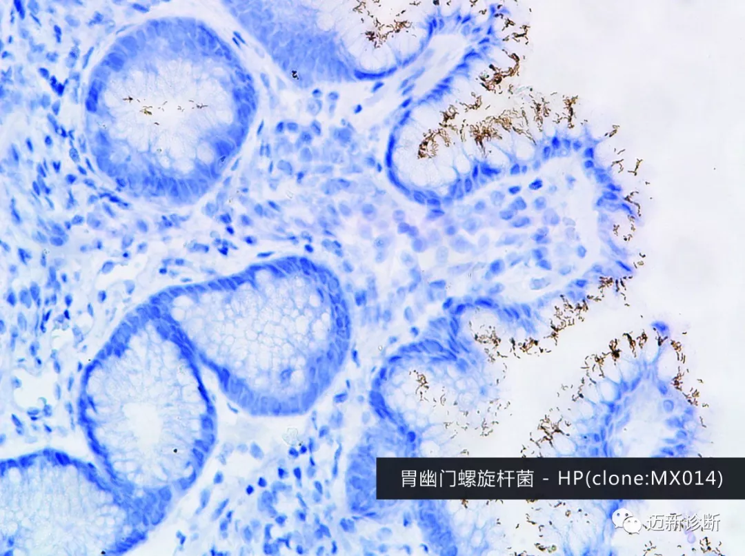
Immunohistochemistry, H. pylori positive


A microbiology expert I often converse with cited an example of a dematiaceous fungal infection, conducting a complete set of examinations from tissue culture, pathology slides, to strain sequencing. This included finding the pathogen in the lesional tissue.He believed that if pathology finds it, then it is definitely meaningful to confirm.This is consistent with the concept I have always advocated of detecting pathogens in situ at the lesion site. I believe,Seeing it on a pathology slide is not enough; one must also be able to recognize it. Due to factors such as slide preparation, sectioning, staining, etc., it may differ from what is seen in microbiology and requires identification and confirmation.Laboratory colleagues believe,Looking for pathogens, searching with purpose will make it easier to find.That is correct. In the pathology department,We usually have two situations: one is searching for pathogens based on clinical clues, and the other is discovering clues of pathogens and then identifying and determining what pathogen it is.At this time, it often requires the assistance of special stains, immunohistochemistry, and even techniques like electron microscopy and molecular methods to aid diagnosis. Providing correct etiological diagnosis should be the direction of our efforts as pathologists.

(Note: Underlined parts are dialogue content from WeChat)

发表回复

您的邮箱地址不会被公开。 必填项已用 * 标注