
Pathology Weekly Reading Notes Episode 5: Immunohistochemical Markers for Diagnosing Epithelial Tumors (Part 2)
Preface:
In previous articles, we provided a detailed compilation and introduction to the CK series among immunohistochemical markers for diagnosing epithelial tumors. However, in practical work, there are many other immunohistochemical markers for diagnosing epithelial tumors, such as mucins, tight junction proteins (Claudin), and certain markers with lineage differentiation characteristics like p63 and p40. Due to space limitations, this issue will focus on introducingmucins and tight junction proteins.,Please stay tuned for other epithelial markers, which will be covered in the next issue.

Immunohistochemical Markers for Diagnosing Epithelial Tumors
II. Mucins
Mucins are a group of high molecular weight, highly glycosylated proteins primarily synthesized by epithelial cells. They consist of approximately 75% carbohydrates and 25% amino acids and can form gel-like substances. Mucins are located on the surface of epithelial cells, functioning as lubricants or acting as chemical barriers for protection. Some mucins are also important components of glandular secretions, such as in salivary glands.
MUC-1
This mucin has many other names, such as the more familiar epithelial membrane antigen (EMA), as well as CD227, Ca15.3, and episialin. It is a transmembrane glycoprotein composed of an intracellular domain and an extracellular domain. This protein is also one of the important components of gastric mucus, protecting the gastric mucosa.
EMA is highly expressed in various types of epithelial cells, mainly glandular epithelium and tumors derived from such epithelium; its expression is very low in squamous cell carcinoma and urothelial carcinoma. Tumor types that are EMA-negative include: basal cell carcinoma, adrenal cortical tumors, malignant melanoma, hepatocellular carcinoma, and germ cell tumors (such as seminoma, embryonal carcinoma, yolk sac tumor).
It should be noted that EMA is not a specific epithelial marker and can be widely expressed in non-epithelial tumors and cell types, such as anaplastic large cell lymphoma, plasma cell tumors, meningioma, epithelioid mesothelioma, perineurioma, synovial sarcoma, epithelioid sarcoma, and neurogenic sarcoma. EMA expression is also commonly seen in nodular lymphocyte-predominant Hodgkin lymphoma.
Since EMA is a highly glycosylated mucin, and some antibodies detect carbohydrate domains, staining results may vary significantly between different antibodies. Overexpression of EMA in carcinoma is associated with poor prognosis.
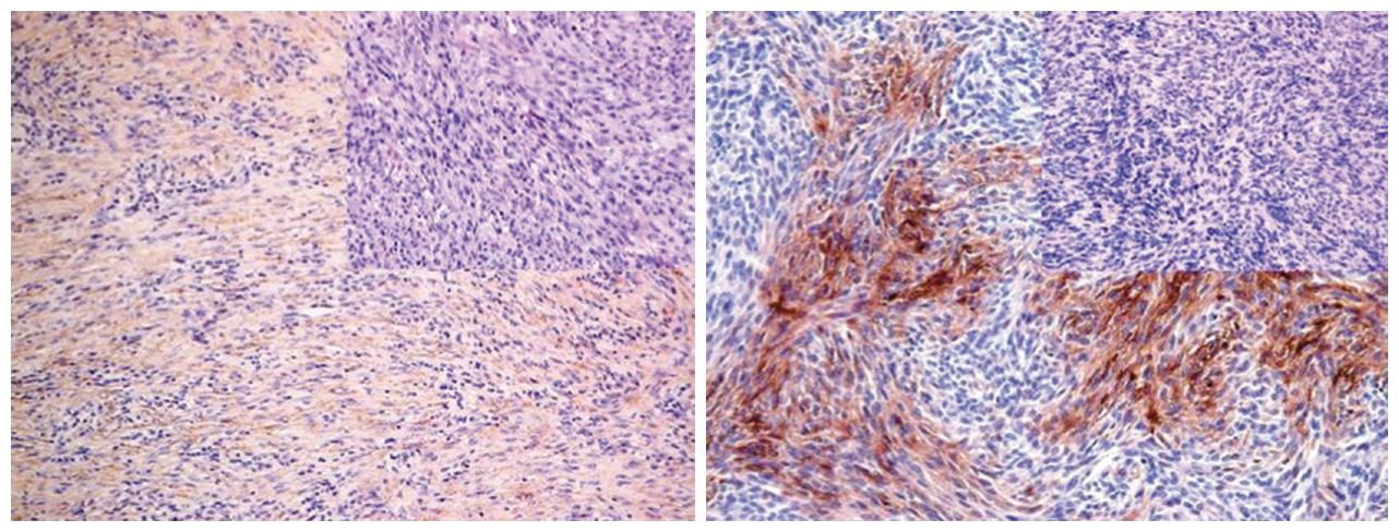
Figure 1. (Left) Positive EMA expression in atypical meningioma; (Right) Focal positive EMA expression in neurogenic sarcoma.
Positive expression pattern: Cell membrane or cytoplasm.
Recommended positive control tissues: Appendix, tonsil.
MUC-2
MUC-3
MUC-4
MUC-5AC
MUC-5B
MUC-6
MUC-16
III. Tight Junction Proteins
Claudins are a group of integral transmembrane proteins, specifically including 23 types. These transmembrane tight junction-associated proteins are found in all cell types with tight junctions, including epithelial and endothelial cells. Claudins form barriers and pores between adjacent cells and regulate molecular transport across intercellular spaces.
In routine immunohistochemical testing, the most commonly used marker is Claudin-4, used to differentiate reactive mesothelial cells from carcinoma cells in pleural and peritoneal effusions. Normally, Claudin-4 is expressed in most epithelial cells and related carcinomas, such as colorectal adenocarcinoma, ovarian cancer, breast cancer, and prostate cancer, while mesothelial cells are negative. Claudin-4 expression can also be seen in endothelial cells, submucosal cells, and the myenteric plexus.
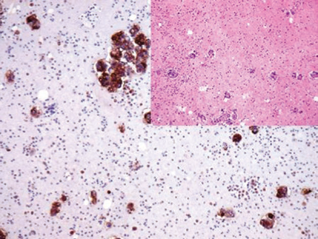
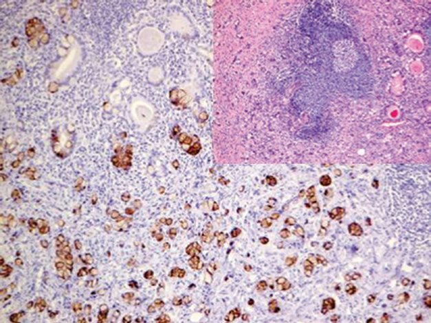
Figure 3. Ovarian cancer cells in ascites, immunohistochemically positive for Claudin-4.
1
Maimai: Currently, more than 15 types of mucins have been identified, and there are as many as 23 types of tight junction proteins. To better select antibodies for clinical pathological diagnosis, we have compiled the following table for reference in the daily work of pathologists.
|
Antibody Name |
Clone Number |
Positive Control |
Positive Location |
|
CA 125* |
MX055 |
Ovarian serous carcinoma, fallopian tube |
Cytoplasm/cell membrane |
|
EMA |
E29 |
Esophageal squamous cell carcinoma, meningioma |
Cytoplasm/cell membrane |
|
MUC-2 |
M53 |
Gastric adenocarcinoma, small intestine tissue |
Cytoplasm |
| MUC-4 | 8G7 | Colon, lung adenocarcinoma | Cytoplasm |
|
MUC5AC |
45M1 | Gastric tissue, gastric adenocarcinoma | Cytoplasm |
| MUC-6 | MRQ-20 | Gastric tissue, gastric adenocarcinoma |
Cytoplasm |
| Claudin-1 | Multipleclones | Colon cancer | CytoMembrane |
*Marked as Maixin clone products
For more information, please contact:800-8581156 or 400-889-9853