
OCCC is a disease whose incidence is related to ethnicity, with significantly higher rates in Asians compared to Caucasians and Africans.It is highly malignant, strongly metastatic, and has a poor prognosis, especially in the middle and advanced stages. Studies have shown thatThe metastasis of OCCC is closely associated with tumor cell subpopulations that have immature and dormant characteristics. However, there are currently no biomarkers that can effectively reflect this subpopulation in OCCC. Therefore, developing new biomarkers is particularly important.
Inhibin α is a glycoprotein kinase belonging to the transforming growth factorβ(TGF-β) superfamily. It is expressed in ovarian granulosa cells, theca cells, testicular Sertoli and interstitial cells, mammary duct and acinar epithelial cells, etc., and is primarily used for the diagnosis of sex cord-stromal tumors.However, its exact role inOCCChas not been reported. Therefore,Shinya Kusumotoand colleagues included2006-2017patients who underwent surgery at Osaka University Hospital between99casesto study the expression characteristics and role ofInhibin α inOCCC.
The results showed thatInhibin α inOCCCcould be detected in both tumor epithelial cells and stromal cells of3Dand2Dcultured cells, it was found thatInhibin α in3Dwas highly expressed inIHCstainingresults indicated that almost allInhibin α-positive cells were negative forKi-67. In3Dcells, the proportion ofInhibin α-positive cells was higher than in2Dcultured cells, and the proportion ofKi-67positive cells was lower than in2Dcultured cells(as shown in1TEXT_32), suggesting the low proliferative characteristics ofInhibin α-expressing cells. Further immunofluorescence double staining and immunoblotting experiments also demonstrated thatInhibin α-expressingOCCCcells are immature.
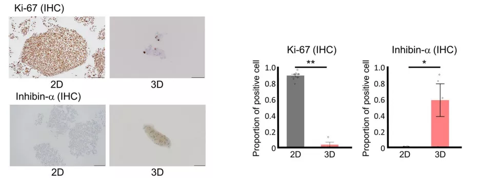
Figure 1: Inhibin α and Ki-67 antibody staining of cells
To validate the above results, the study performed immunoblotting and ALDEFLUOR analysis for Inhibin α in OCCC cell lines with high and low aldehyde dehydrogenase levels. It was found that Inhibin α levels were higher in the high aldehyde dehydrogenase population than in the low aldehyde dehydrogenase population. In the publicly available microarray dataset GSE129617, consisting of 25 OCCC patients, Inhibin α positivity was positively correlated with stem cell genes (Sox2, Klf4, Oct 3/4) and negatively correlated with the proliferation-related gene MKI67. In five pathologically diagnosed recurrent cases, the H-score of Inhibin α at the recurrence site was higher than at the primary site (as shown in Figure 2). These data further demonstrate that Inhibin α-expressing OCCC cells are in an undifferentiated, immature state. Therefore, Inhibin α can serve as an effective biomarker for immature cell subpopulations in OCCC.
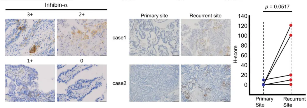
Figure 2: Inhibin α staining of OCCC tissue sections at initial diagnosis and recurrence
(H-score calculated based on Inhibin α IHC staining of OCCC tissues with high (3 points), medium (2 points), low (1 point), or zero (0 points) signal intensity)
When studying the characteristics of Inhibin α-expressing OCCC cells, it was found in the publicly available microarray datasets GSE129617 and GSE29450, which include 10 OCCC patients, that Inhibin α was positively correlated with VEGF-A and VEGF-C (as shown in Figure 3A). Moreover, VEGF-A and VEGF-C expression was higher in Inhibin α-positive cells than in Inhibin α-negative cells. This indicates that Inhibin α-expressing OCCC cells have high angiogenic potential, which may increase the probability of metastasis/recurrence. Further immunoblotting also confirmed this conclusion.
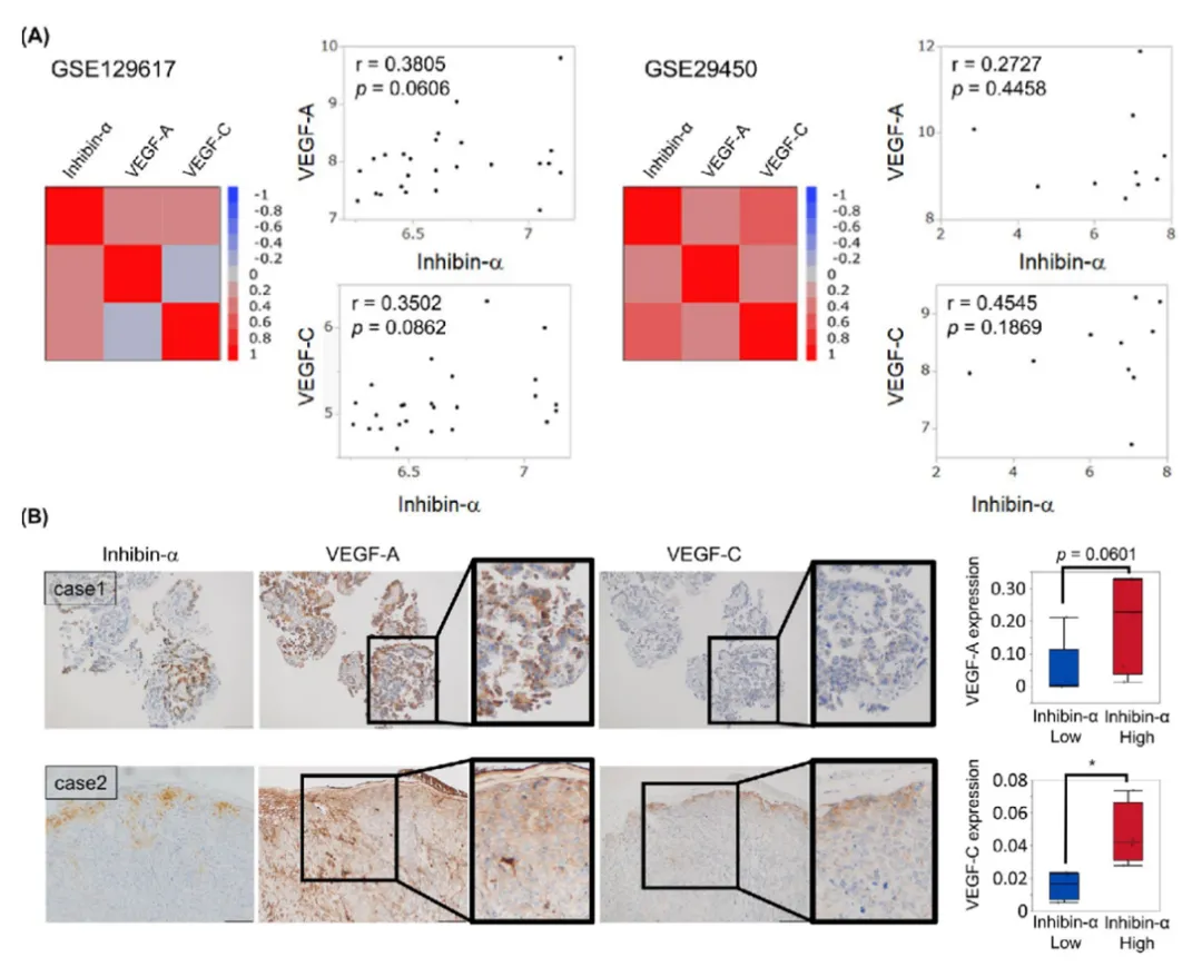
Figure 3: Inhibin α-positive cells have higher angiogenic potential
(A: Correlation study of Inhibin α, VEGF-A, and VEGF-C in GSE129617 and GSE29450; Representative IHC images of Inhibin α, VEGF-A, and VEGF-C in OCCC tissues)
Additionally, the study conducted a follow-up on 69 OCCC patients in the sample (35 Inhibin α-positive, accounting for 51%; 34 Inhibin α-negative, accounting for 49%). It was found that compared to the Inhibin α-negative group, patients in the Inhibin α-positive group had significantly lower OS and DFS. There were 8 deaths and 14 cases of progressive disease in the Inhibin α-positive group, versus 1 death and 3 cases of progressive disease in the Inhibin α-negative group. Kaplan–Meier plots showed substantial separation of survival curves for OS and DFS (OS: p=0.012, HR=9.06, 95% CI: 1.31–72.56; DFS: p=0.001, HR=6.02, 95% CI: 1.73-20.97, as shown in Figure 4B). Cox proportional hazards model showed that positive Inhibin α expression was significantly positively correlated with poor prognosis (OS: p=0.0086, HR=9.03, 95% CI: 1.63-168.61; DFS: p=0.0011, HR=6.02, 95% CI: 1.95-26.26), even after adjusting for stage. In IPW analysis, after adjusting for confounding factors, the concordance index was 0.640. IPW analysis and propensity scores indicated that positive Inhibin α expression had a significant impact on patient prognosis, leading to poor outcomes (as shown in Figure 4C).
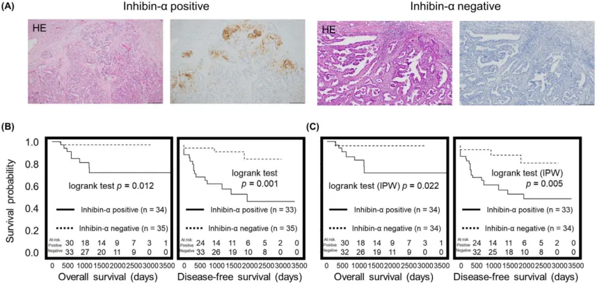
Figure 4: Evaluation of OS and DFS in Inhibin α-positive or negative cases
(A) Typical images of Inhibin α immunohistochemistry; (B) Kaplan-Meier plot; (C) IPW analysis plot
Based on the above research, it was found that Inhibin α is expressed in low-proliferating OCCC cells. These cells have immature characteristics and high angiogenic potential, which may increase the likelihood of metastasis/recurrence, significantly impact recurrence risk and prognosis, and lead to poor outcomes. These findings indicate that in OCCC, Inhibin α is a biomarker for immature cells and also an independent prognostic biomarker. Therefore, it may become important new information for determining treatment strategies.
|
Antibody Name
|
Product Number
|
Clone Number
|
Cellular Localization
|
|
Inhibin α
|
MAB-0891
|
MX098
|
Cytoplasm
|
References:
[1] Kusumoto Shinya,Kurashige Masako,Ohshima Kenji,et al. An immature inhibin-α-expressing subpopulation of ovarian clear cell carcinoma cells is related to an unfavorable prognosis. [J]. Cancer medicine2021,10(5):1485-1500.
