
Reading Literature Together: H3.3 G34W as a Reliable Marker for Diagnosing and Differentiating Giant Cell Tumor of Bone
Introduction:
Materials and Methods:
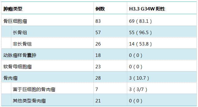
Research Results:
|
Number of Tumors |
Staining Results |
|||
|
Tumor Type |
Total |
Positive |
Negative |
Positive Rate |
|
Giant Cell Tumor of Bone |
136 |
115 |
17 |
87.5% |
|
Chondroblastoma |
34 |
— |
34 |
|
|
Aneurysmal Bone Cyst |
20 |
— |
20 |
|
|
Tenosynovial Giant Cell Tumor |
29 |
— |
29 |
|
|
Brown Tumor |
3 |
— |
3 |
|
|
Non-Ossifying Fibroma |
19 |
— |
19 |
|
|
Giant Cell-Rich Osteosarcoma |
3 |
— |
3 |
|
|
Other Types of Osteosarcoma |
31 |
1 |
30 |
3.2% |
|
Tumor Type |
Number of Cases |
Positive |
Negative |
False Positive Rate |
False Negative Rate |
|
Giant Cell Tumor of Bone |
136 |
119 |
17 |
0.7% |
12.5% |
|
Non-Giant Cell Tumor of Bone |
139 |
1 |
138 |
||
|
Sensitivity |
— |
87.5% |
— |
— |
|
|
Specificity |
— |
99.3% |
— |
— |
|
The results from Teacher Wang Xuan’s article showed that the sensitivity of H3.3 G34W immunohistochemistry for diagnosing giant cell tumor of bone was 83.1%, and the specificity was 95.7%.
Diagnostic Image Appreciation:
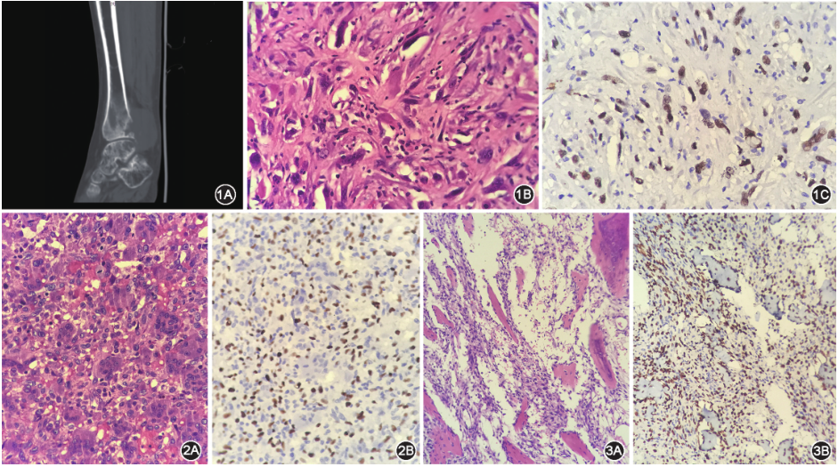
Figure 1 Osteosarcoma with positive H3F3A G34W immunohistochemistry;Figure A shows imaging data;Figure B shows tumor cells are spindle-shaped, with large, hyperchromatic nuclei, visible nucleoli, and obvious atypia, HE high magnification;Figure C shows tumor cells exhibit strong nuclear positivity for H3F3A G34W immunohistochemical staining, Lumatas (Titan) fullyautomated immunohistochemical staining, high magnification;Figure 2 Giant cell tumor of bone;Figure A shows neoplastic mononuclear stromal cells with round or oval nuclei, indistinct borders, and evenly distributed osteoclast-like giant cells among the mononuclear stromal cells, HE medium magnification;Figure B shows neoplastic mononuclear stromal cells exhibit strong nuclear positivity for H3F3A G34W immunohistochemical staining, while osteoclast-like giant cell nuclei are negative, Lumatas (Titan) fully automated immunohistochemical staining, medium magnification;Figure 3 Giant cell tumor of bone after denosumab treatment;Figure A shows spindle cell proliferation, significant disappearance of osteoclast-like giant cells, and reactive bone hyperplasia, HE medium magnification;Figure B shows spindle cells between reactive bone exhibit strong nuclear positivity for H3F3A G34W immunohistochemical staining, Lumatas (Titan) fully automated immunohistochemical staining, medium magnification
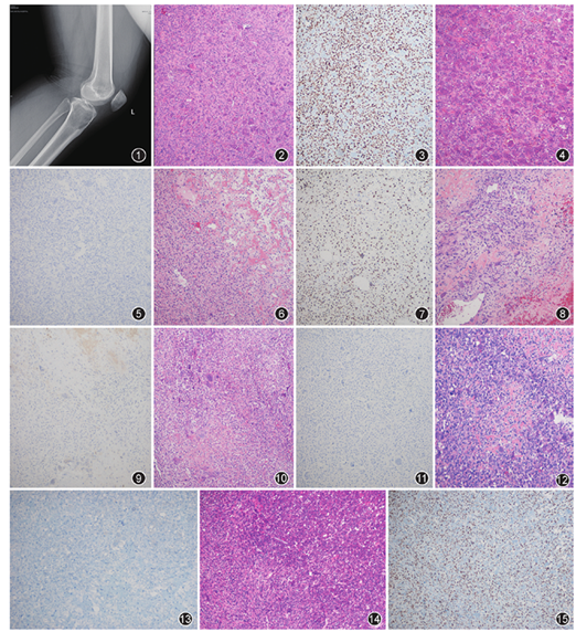
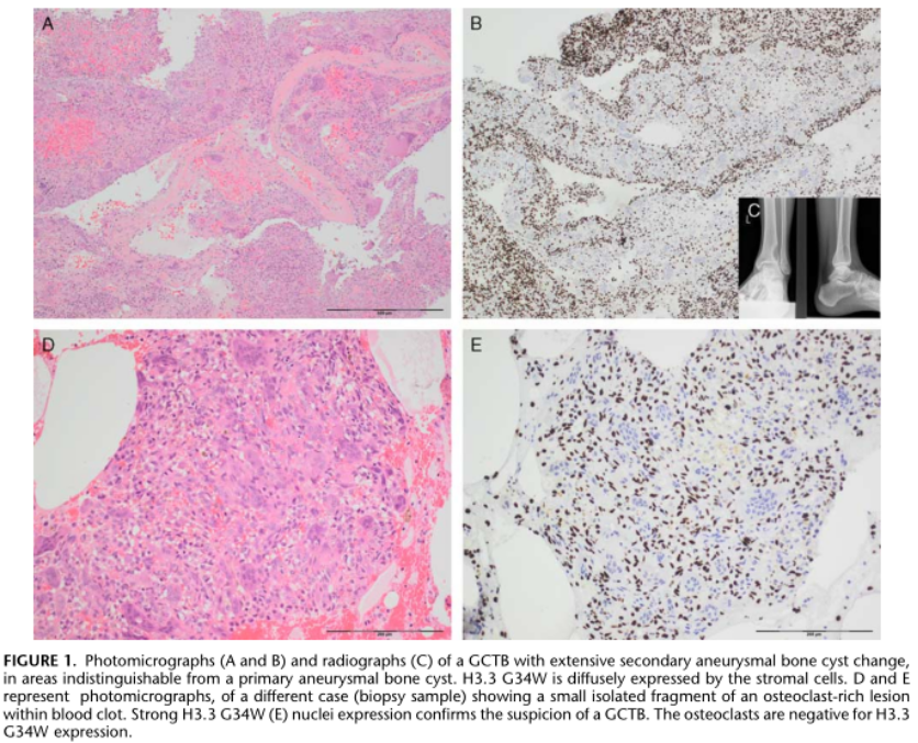
Figure 1 Giant cell tumor of bone (GCTB) in the upper end of the left tibia, X-ray shows expansive bone destruction with relatively clear boundaries and no sclerotic band;Figure 2 GCTB occurring in long bones, composed of sheets of oval mononuclear tumor cells and evenly distributed osteoclast-like giant cells, HE low magnification;Figure 3 H3.3 G34W staining in GCTB occurring in long bones is only seen in the nuclei of mononuclear tumor cells, showing diffuse strong positive expression, EnVision method, low magnification;Figure 4 GCTB occurring in non-long bones, composed of sheets of oval mononuclear tumor cells and evenly distributed osteoclast-like giant cells, microscopic morphology is basically consistent with GCTB occurring in long bones, HE low magnification;Figure 5 H3.3 G34W negative in GCTB occurring in non-long bones, EnVision method, low magnification;Figure 6 GCTB after denosumab treatment shows spindle cell proliferation with varying degrees of fibrosis or new bone formation, osteoclast-like giant cells are reduced or even disappeared, HE low magnification;Figure 7 Spindle cells in GCTB after denosumab treatment show H3.3 G34W nuclear positivity, and H3.3 G34W expression is observed in immature osteoblasts, EnVision method, low magnification;Figure 8 Aneurysmal bone cyst, showing cystic wall-like tissue, stroma rich in fibroblasts, and visible osteoclast-like giant cells and reactive new bone, HE low magnification;Figure 9 H3.3 G34W negative in aneurysmal bone cyst, EnVision method, low magnification;Figure 10 Chondroblastoma, oval mononuclear tumor cells diffusely distributed, with many osteoclast-like giant cells, occasional chicken-wire calcification, HE low magnification;Figure 11 H3.3 G34W negative in chondroblastoma, EnVision method, low magnification;Figure 12 Giant cell-rich osteosarcoma (GCRO), mononuclear tumor cells are oval or spindle-shaped, with atypia, pathological mitotic figures visible, focal tumor osteoid formation, stroma shows numerous reactive osteoclast-like giant cells, HE low magnification;Figure 13 H3.3 G34W negative in GCRO, EnVision method, low magnification;Figure 14 Primary malignant GCTB, tumor tissue contains typical giant cell tumor components, and the boundary with spindle cell sarcoma components is clear, HE low magnification;Figure 15 Both benign and malignant components in primary malignant GCTB show H3.3 G34W nuclear positive expression in mononuclear stromal cells, EnVision method, low magnification
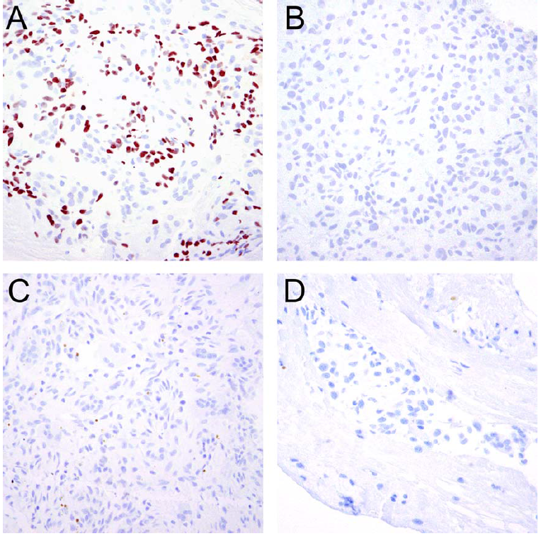
Immunohistochemistry for H3G34W in (A) a giant cell tumor of bone was positive in a subset of mononuclear tumorcells (approximately 40% of cells) and negative in multinucleated giant cells as well as the remaining mononuclear cells and inflammatory cells. H3G34W was negative in (B) chondroblastoma, (C) aneurysmal bone cyst, and (D) osteosarcoma.
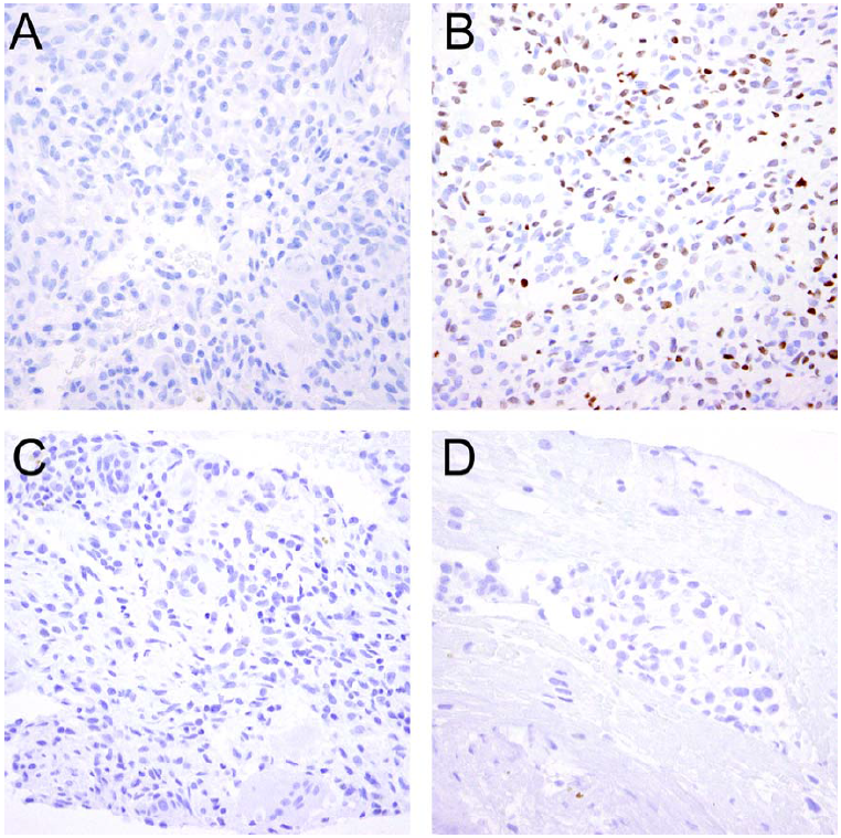
Immunohistochemistry for H3K36M was negative in (A) giant cell tumor of bone, but positive in (B) the neoplastic cells of chondroblastoma. (C) Aneurysmal bone cyst and (D) osteosarcoma were negative for H3K36M.
The H3.3 G34W antibody is a highly sensitive and specific biological marker for diagnosing giant cell tumor of bone, aiding in differentiation from bone tumors with similar morphology (such as aneurysmal bone cyst, chondroblastoma, tenosynovial giant cell tumor, and osteosarcoma). Its use in routine work is recommended.
-
H3.3 G34W expression is only seen in mononuclear cells; osteoclast-like giant cells, foam histiocytes, lymphocytes, endothelial cells, vascular smooth muscle cells, and fibroblasts show no expression.
-
H3.3 G34W is a specific biological marker for giant cell tumor of bone and is not expressed in tumors with similar morphology such as aneurysmal bone cyst, chondroblastoma, tenosynovial giant cell tumor, and brown tumor.
-
In cases of giant cell-rich osteosarcoma, H3.3 G34W immunohistochemical staining can be positive or negative, consistent with previous studies. Therefore, differential diagnosis between giant cell-rich osteosarcoma and giant cell tumor of bone requires more attention to tumor cell atypia, pathological mitotic figures, and tumor osteoid tissue.
-
Chondroblastoma does not have H3F3A G34 site mutations; most mutations occur at the H3F3B K36 site.
-
H3.3 G34W immunohistochemistry aids in the differential diagnosis of giant cell tumor of bone after denosumab treatment from other tumors with similar morphology.
-
A very small portion of giant cell tumors of bone are negative for H3.3 G34W immunohistochemistry, and the positive rate in the non-long bone group is significantly lower. H3.3 G34W site mutations most commonly occur in long bones, while rare subtypes such as H3.3 p.G34L, M, V often occur in hand bones, patella, and axial bones. If these sites are excluded, the positive expression rate of H3.3 p.G34W antibody in GCTB is as high as 97.8%.
Maixin Related Antibody Information:
|
Antibody Name:H3.3 G34W |
||
|
Product Number: RMA-0810 |
Clone Number: RM263 |
Rabbit Anti-Human: Monoclonal Antibody |
|
Applicable Tissue: Paraffin Sections |
Positive Location: Nucleus |
Pretreatment: EDTA Heat Retrieval |
Source Literature:
[1] Wang Xuan, Wu Nan, Zhang Rusong, et al. Expression and Diagnostic Value of H3.3 G34W Mutant Antibody in Giant Cell Tumor of Bone [J]. Chinese Journal of Pathology, 2020, 49(02): 116-121. DOI: 10.3760/cma.j.issn.0529-5807.2020.02.003
[2] Bai Yueqing, Yang Tingting, Zhang Huizhen. Application Value of H3F3A G34W Immunohistochemical Staining in the Diagnosis of Giant Cell Tumor of Bone [J]. Chinese Journal of Pathology, 2019, 48(7): 531-536. DOI: 10.3760/cma.j.issn.0529-5807.2019.07.006
【3】Amary F, Berisha F, Ye H et al,H3F3A (Histone 3.3) G34W immunohistochemistry: a reliable marker defining benign and malignant giant cell tumor of bone. Am J Surg Pathol 41(8):1059–1068
【4】Schaefer, I.‐M., Fletcher, J.A., Nielsen, G.P., Shih, A.R., Ferrone, M.L., Hornick, J.L. and Qian, X., Immunohistochemistry for histone H3G34W and H3K36M is highly specific for giant cell tumor of bone and chondroblastoma, respectively, in FNA and core needle biopsy. Cancer Cytopathology, 126: 552-566. doi:10.1002/cncy.22000