Tp53The protein normally expressed by the gene is normal/wild-type p53, which is expressed in the nuclei of all normal cells. Due to its short half-life of only 10-20 minutes, it is not easily detectable by immunohistochemistry. Normal/wild-type p53 protein binds to DNA and negatively regulates cell growth and division. In the case of DNA damage, normal/wild-type p53 halts the cell cycle until repair occurs. If repair is not possible, normal/wild-type p53 induces apoptosis.
Tp53Gene mutations are associated with the occurrence and prognosis of various cancers.Tp53The protein expressed after gene mutation is mutant p53.TP53The most commonly used method for gene mutation analysis is the detection of mutant p53 by immunohistochemistry, which is rapid and easy to perform. Mutant p53 loses its inhibitory effect on cell proliferation, allowing damaged and abnormal cells to proliferate uncontrollably, ultimately leading to tumor formation. Mutant p53 not only serves as a poor prognostic indicator for various tumors but also assists in the differential diagnosis of malignant lesions (carcinoma in situ vs. invasive carcinoma) and reactive lesions, as well as in distinguishing between serous and endometrioid histological types, predicting outcomes within a given histological type, or predicting outcomes across multiple histological types.
p53 “Positive” Expression Patterns
Early studies found that nonsynonymousTp53gene missense mutations lead to nuclear accumulation of p53 protein, which can be detected as p53 protein overexpression by immunohistochemistry. p53 protein expression manifests as a diffuse, strong nuclear positivity involving at least 80% of tumor cells, but typically nearly 100%. With continuous improvements in immunohistochemistry, other abnormal p53 expression patterns associated with the presence ofTp53gene mutations have been observed. Therefore, p53 staining should not be simply reported as positive or negative. The staining pattern should be interpreted as wild-type or abnormal/mutant-type, with abnormal/mutant-type including overexpression, complete absence, and cytoplasmic expression. An abnormal p53 staining pattern almost certainly indicates a potentialTp53gene mutation. The staining patterns are shown in Figure 1.
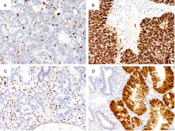
Figure 1. Different patterns of p53 expression. (A) Endometrioid carcinoma showing a normal wild-type p53 expression pattern, with varying proportions and intensities of nuclear staining in tumor cells. (B) Overexpression in a grade 3 endometrioid carcinoma, showing strong staining in almost all tumor cell nuclei, stronger compared to the internal control of central fibroblasts. (C) Endometrial serous carcinoma showing complete absence of p53 expression, with internal controls showing moderate to strong but variable staining. (D) Endometrioid carcinoma showing cytoplasmic p53 expression, with internal controls (stroma and normal endometrial glands) showing a nuclear wild-type pattern. The cytoplasmic pattern is accompanied by nuclear staining of similar intensity.
Wild-type p53 Staining Pattern
The normal wild-type pattern shows a significant range of staining, from only a few positive tumor cell nuclei to most nuclei being positive (as shown in Figure 1.A). The expression level of wild-type p53 depends on the differentiation state of the cell and is related to proliferative activity. Tumors with a higher proliferation index typically show more p53 staining, and tumors with so-called “high” wild-type staining may be confused with overexpression.
It is noteworthy that some splice site mutations or truncating mutations (the latter characterized by C-terminal stop gains) result in non-functional p53 protein detected by IHC, with a staining pattern that appears as the normal wild-type pattern.
Abnormal/Mutant-type — Overexpression Staining Pattern
NonsynonymousTp53gene missense mutations lead to nuclear accumulation of p53 protein, which can be detected as p53 protein overexpression by immunohistochemistry. Strong staining is observed in almost all tumor cell nuclei, with tumor cell nuclei staining stronger than the internal control central fibroblasts (as shown in Figure 1.B). The distinction between overexpression and wild-type primarily involves observing whether tumor cells contain negative, weakly positive, and strongly positive cells. The presence of tumor cells with these three different staining intensities is interpreted as a wild-type staining pattern, whereas tumor cells that are almost entirely positive are interpreted as the overexpression staining pattern within the abnormal/mutant-type category.
Abnormal/Mutant-type — Complete Absence Staining Pattern
When theTp53gene undergoes a loss-of-function mutation, the mutant p53 staining pattern detected by IHC is the complete absence pattern within the abnormal/mutant-type category (as shown in Figure 1.C). Before interpreting p53 staining as abnormal complete absence, adequate staining of internal controls (fibroblasts, endothelial cells, or lymphocytes) must be observed. Cases without positive internal controls are uninterpretable. The distinction between complete absence and wild-type involves observing whether the internal positive control is valid and whether tumor cells exhibit weakly positive, strongly positive, and negative cells.
Abnormal/Mutant-type — Cytoplasmic Staining Pattern
Through optimization of immunohistochemistry, a fourth, less common p53 staining pattern has been observed. This cytoplasmic pattern is characterized by distinct cytoplasmic staining accompanied by variable nuclear staining (as shown in Figure 1.D and Figure 2). Excessively high primary antibody concentration may show strong diffuse nuclear staining with low-intensity cytoplasmic background, which should be interpreted as overexpression rather than a cytoplasmic pattern. In tubo-ovarian high-grade serous carcinoma, the cytoplasmic pattern is associated with mutations that disrupt the nuclear localization domain of the p53 protein. Distinguishing wild-type from the cytoplasmic pattern primarily involves observing cytoplasmic staining intensity, whether there are clearly defined weakly positive, strongly positive, and negative cells, and whether cytoplasmic staining is similar in intensity to nuclear staining.
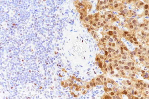
Figure 2. p53 cytoplasmic expression pattern, with cytoplasmic expression accompanied by nuclear staining of similar intensity. Lymphocytes in the image are positive.
p53 plays a crucial role in tumor diagnosis, classification, and prognosis prediction. Accurate interpretation and effective immunohistochemistry staining protocols are prerequisites for the correct interpretation of p53 results. The application of p53 immunohistochemistry in gynecological tumors will be detailed in the following article. Please stay tuned.
References
1. M Köbel, Piskorz AM, Lee S, et al. Optimized p53 immunohistochemistry is an accurate predictor of TP53 mutation in ovarian carcinoma[J]. Journal of Pathology Clinical Research, 2016, 2(4):247-258.
2. M Köbel, BM Ronnett, N Singh, et al. Interpre-tation of P53 Immunohistochemistry in Endometrial Carcinomas: Toward Increased Reproducibility[J]. International Journal of Gynecological Pathology, 2018:1.
