
Overview of Immunohistochemistry for Skeletal Muscle Tumors
Commonly used immunohistochemical markers for the diagnosis of skeletal muscle tumors include Desmin, Myoglobin, Myogenin, Myosin, MyoD1, EGFR, fibrillin-2, and p-cadherin.
Table 1. Commonly Used Immunohistochemical Markers for Diagnosing Skeletal Muscle Tumors
(Click to view larger image)
Note:
-
A small portion of differentiated rhabdomyosarcomas show varying degrees of CK expression, which may lead to misdiagnosis.
Detailed Explanation of Some Markers
Positive Expression Pattern: Cytoplasm
Recommended Positive Control Tissue: Appendix
Desmin is a type III intermediate filament protein expressed in the intercalated discs and Z-lines of cardiac muscle and the Z-lines of skeletal muscle. Therefore, this marker can be used to label cardiac muscle, skeletal muscle, smooth muscle, and tumors arising from these cells. The degree of positivity for this marker is related to the differentiation level of the corresponding muscle cells or tumors.
In clinical practice, Desmin is an important diagnostic marker for all myogenic tumors and tumors with myogenic differentiation, while myoepithelial cells are negative. However, other tumors morphologically similar to rhabdomyosarcoma can also show positive expression of this marker, such as desmoplastic small round cell tumor and alveolar soft part sarcoma. Therefore, immunohistochemical testing for rhabdomyosarcoma should include at least one myogenic transcription regulatory protein (Myogenin, MyoD1); markers for smooth muscle differentiation should also be added. It is also important to note that mesothelioma (mainly the sarcomatoid subtype) and, in extremely rare cases, carcinoma can show focal Desmin positivity. Therefore, for difficult cases, CK testing should be added.
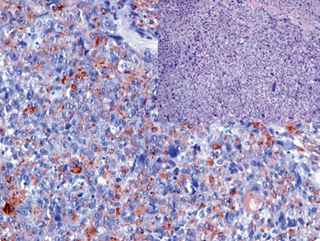
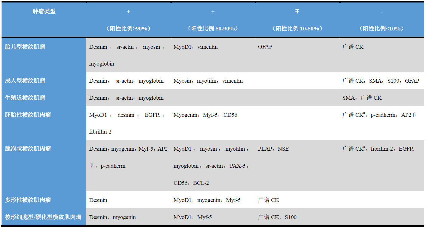
Figure 1. Pleomorphic rhabdomyosarcoma, immunohistochemistry shows significant expression of desmin in the cytoplasm of tumor cells.
Positive Expression Pattern: Cytoplasm
Recommended Positive Control Tissue: Skeletal Muscle
Myoglobin is an iron-binding, oxygen-binding single-chain polypeptide that appears in the early stages of muscle tissue differentiation. This marker is expressed in skeletal muscle, cardiac muscle, rhabdomyoblasts, and adult-type skeletal muscle tumors. Embryonal muscle tumors, smooth muscle tumors, and other sarcomas do not express Myoglobin. However, according to reports, several carcinomas show weak to moderate expression of this marker, specifically breast cancer, prostate cancer, colon cancer, and head and neck cancers, which may be related to hypoxia and steroid hormone receptor positivity.
Positive Expression Pattern: Nucleus
Recommended Positive Control Tissue: Rhabdomyosarcoma/Fetal Muscle Tissue
The MyoD family of myogenic transcription regulatory factors includes MyoD1 (Myf-3), myogenin (Myf-4), Myf-5, and MRF-4 (Myf-6). These transcription factors are involved in the activation of muscle stem cells and participate in the regulation of early embryonic skeletal muscle differentiation, the maintenance of myogenic programs, and muscle repair. The expression of MyoD1 and myogenin is downregulated in mature skeletal muscle, and these two markers are expressed only in all rhabdomyosarcoma-type cells.
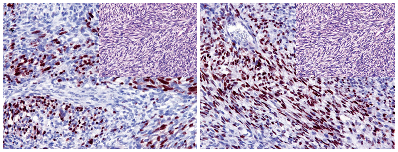
Figure 2. In rhabdomyosarcoma, Myogenin (left) and MyoD1 (right) show strong positive nuclear expression.
It should be noted that both of these myogenic markers can be positive in non-neoplastic myoblasts, which can be seen in reparative and atrophic muscle lesions. Expression of Myogenin and MyoD1 has also been reported in certain other tumors, such as desmoid tumor, infantile fibrosarcoma, and Wilms tumor. In the interpretation of Myogenin and MyoD1 staining, only nuclear staining should be considered positive; other staining patterns (such as cytoplasmic or membranous staining) are diagnostically insignificant artifacts.
Pax-5 has been described in B-lymphocyte markers and is also a marker for some neuroendocrine carcinomas. For non-lymphocytic tumors, Pax-5 can be expressed in alveolar rhabdomyosarcoma but is generally negative in embryonal rhabdomyosarcoma.
EGFR is a type 1 receptor tyrosine kinase, which has been described in previous sections. EGFR is a transmembrane glycoprotein normally expressed on the cell membrane of various normal epithelial and non-epithelial cells. Therefore, its expression is a characteristic indicator for various epithelial and non-epithelial tumors and is an indicator for differentiating embryonal rhabdomyosarcoma from other types of rhabdomyosarcoma.
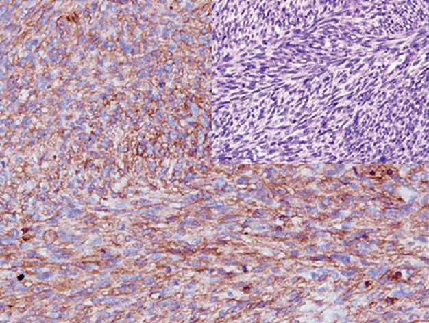
Figure 3. In embryonal rhabdomyosarcoma, EGFR shows strong positive expression.
Overview of Immunohistochemistry for Smooth Muscle Tumors
Commonly used immunohistochemical markers for the diagnosis of smooth muscle tumors include Desmin, SMA, h-caldesmon, Calponin, Smoothelin, Transgelin, and steroid hormone receptors.
Table 2. Commonly Used Immunohistochemical Markers for Diagnosing Smooth Muscle Tumors
(Click to view larger image)
Detailed Explanation of Some Markers
Positive Expression Pattern: Cytoplasm
Recommended Positive Control Tissue: Appendix
Smooth muscle actin (SMA) is a type of actin. Actin is a major cytoskeletal protein, a group of contractile microfilaments including α, β, and γ subunits. α-actin has three isoforms: type 1 is cardiac muscle actin, type 2 is smooth muscle actin, and type 3 is skeletal muscle actin (sr-actin). In fact, type 2 α-actin antibodies can label smooth muscle cells, myoepithelial cells, and myofibroblasts. The actin clone 1A4 is widely used as a marker for smooth muscle actin and is effective for diagnosing smooth muscle, myoepithelial, and myofibroblastic lesions. Another widely used actin clone is HHF-35, which reacts with both skeletal muscle actin and smooth muscle actin, and thus can be used for staining smooth muscle tumors and skeletal muscle tumors.
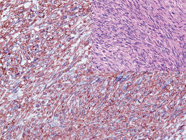
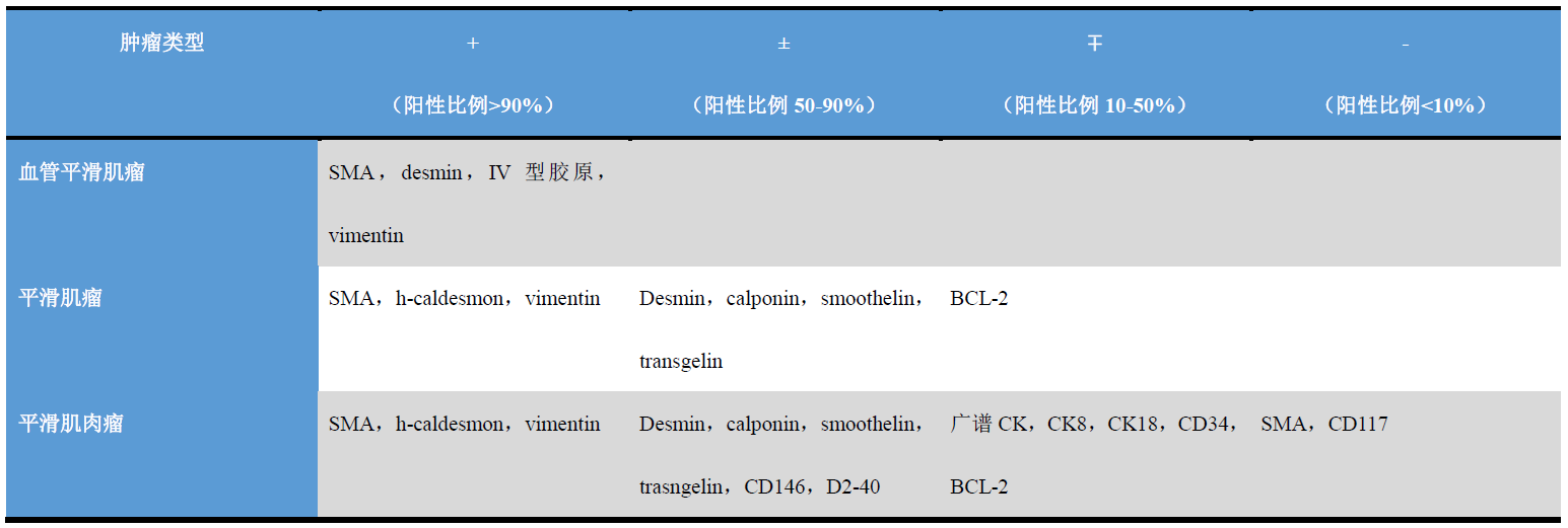
Figure 4. In leiomyosarcoma, SMA shows strong positive cytoplasmic expression.
It should be noted that SMA expression can be seen in certain other types of tumors morphologically similar to smooth muscle tumors, such as endometrial stromal tumors, synovial sarcoma, GIST, and sarcomatoid mesothelioma.
Positive Expression Pattern: Cytoplasm
Recommended Positive Control Tissue: Appendix
Caldesmon is an intracellular calmodulin-binding protein involved in the regulation of smooth muscle contraction. Caldesmon has two isoforms – a low molecular weight isoform (l-Caldesmon), involved in modulating the cytoskeleton and cell morphology and regulating cell proliferation; and a high molecular weight isoform (H-Caldesmon), mainly expressed in visceral and vascular smooth muscle cells and myoepithelial cells. In routine histopathology, because H-Caldesmon expression in non-smooth muscle tumors is less common than SMA, this marker is used as a specific marker for smooth muscle tumors; and unlike Actin, myofibroblasts do not express H-Caldesmon.
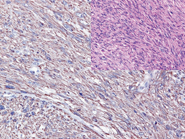
Figure 5. Leiomyosarcoma, showing strong positive cytoplasmic expression of H-Caldesmon.
However, H-Caldesmon can also show positive expression in certain non-smooth muscle lesions, such as gastrointestinal stromal tumors, inflammatory myofibroblastic tumor, and pleural and peritoneal epithelioid mesothelioma, which should be noted in differential diagnosis.
Positive Expression Pattern: Cytoplasm
Recommended Positive Control Tissue: Appendix
Calponin is a cytoskeleton-associated protein involved in the regulation of smooth muscle contraction. The expression specificity of this marker is similar to H-Caldesmon, but generally GIST does not express Calponin.
Transgelin is an actin-binding gel protein of the Calponin family, expressed in the cytoplasm and cell membrane of smooth muscle cells. Transgelin is the earliest marker of smooth muscle differentiation and can be expressed in visceral and vascular smooth muscle cells, myofibroblasts, and related benign and malignant tumors. This marker can also be expressed in epithelioid tumor cells in basal-like triple-negative breast cancer and in epithelioid tumor cells in a small portion of malignant nerve sheath tumors. Rhabdomyosarcoma, GIST, and endometrial stromal tumors do not express it.
Transgelin can also be expressed in fibroblasts, myofibroblasts, and some epithelial cells.
Smoothelin is a component of the cytoskeleton in differentiated smooth muscle cells. This protein has two isoforms: type A, composed of short chains, expressed in visceral smooth muscle; type B, composed of long chains, unique to vascular smooth muscle. Myoepithelial cells, myofibroblasts, skeletal muscle, and cardiac muscle do not express this marker. Smoothelin is a specific marker for smooth muscle tumors, and its expression is related to the differentiation grade of these tumors.
Figure 6. Leiomyosarcoma, tumor cells express Smoothelin.
This marker can also be used to display the muscularis mucosae and muscularis propria in the evaluation of bladder and intestinal tumors; however, for intestinal tumors, it is recommended to use it in conjunction with SMA for comparison.
MaiMai Recommendation: “Factors affecting immunohistochemistry results are diverse, but high-quality antibodies will be an effective guarantee for correct diagnosis. This issue of MaiMai recommends the following antibodies, you might want to try them.””
|
Antibody Name
|
Clone Number
|
Positive Control
|
Cellular Localization
|
|
Calponin*
|
MX023
|
Breast tissue, leiomyoma
|
Cytoplasm
|
|
Desmin*
|
MX046
|
Esophageal tissue, appendix
|
Cytoplasm
|
|
Myo D1*
|
MX049
|
Rhabdomyosarcoma
|
Nucleus
|
|
Myogenin*
|
MX078
|
Rhabdomyosarcoma
|
Nucleus
|
|
Myoglobin
|
Polyclonal
|
Striated muscle
|
Cytoplasm
|
|
SMA
|
1A4
|
Colonic smooth muscle, appendix
|
Cytoplasm
|
*Marked as Maxin clone products
For more information, please contact: 800-8581156 or 400-889-9853
