
Common Immunohistochemical Markers for Malignant Melanoma
Commonly used immunohistochemical markers for diagnosing malignant melanoma include HMB45,Melan A, tyrosinase, SOX-10, microphthalmia transcription factor (MiTF), WT-1, S100, CD63 (NK-C3), PHH3, Ki-67.
Table 1. Overview of Common Immunohistochemical Markers for Malignant Melanoma
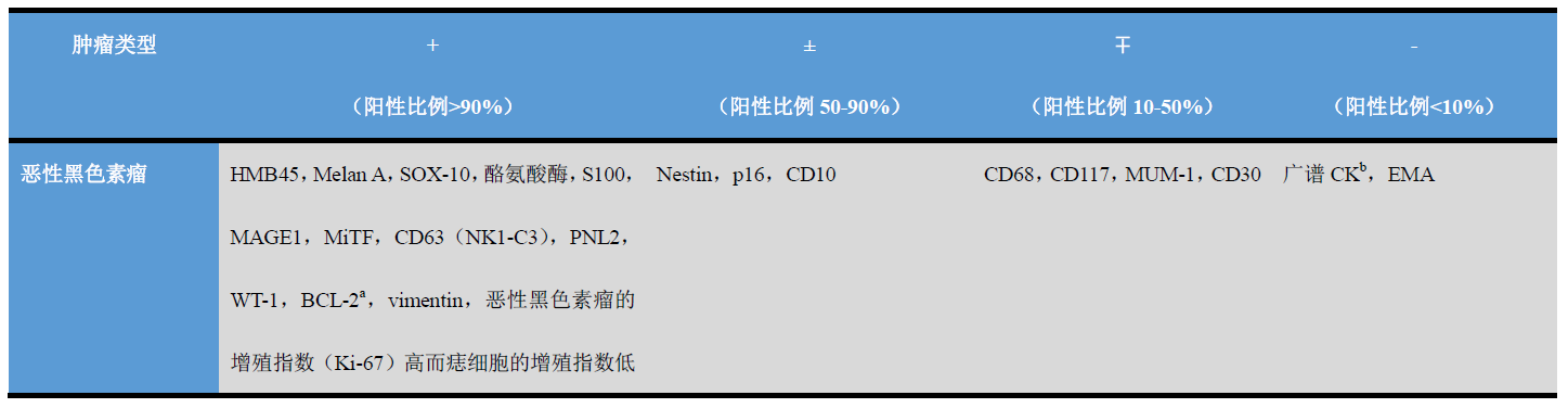
(Click to view larger image)
Note:
-
In benign nevi, it is generally negative;
-
In a small number of malignant melanomas, there may be focal weak positive expression of CK.
Detailed Explanation of Some Markers
Positive expression pattern: Cytoplasm
Recommended positive control tissue: Malignant melanoma
HMB45 is a glycoprotein associated with the maturation of melanosomes from stage I to stage II. In normal tissues, HMB45 can be expressed in retinal pigment epithelium and fetal melanocytes, but not in mature melanocytes or intradermal nevi. HMB45 is a marker for melanocytic tumors and tumors with melanocytic differentiation, such as various types of malignant melanoma, desmoplastic nevus, Spitz nevus, blue nevus, and clear cell sarcoma.
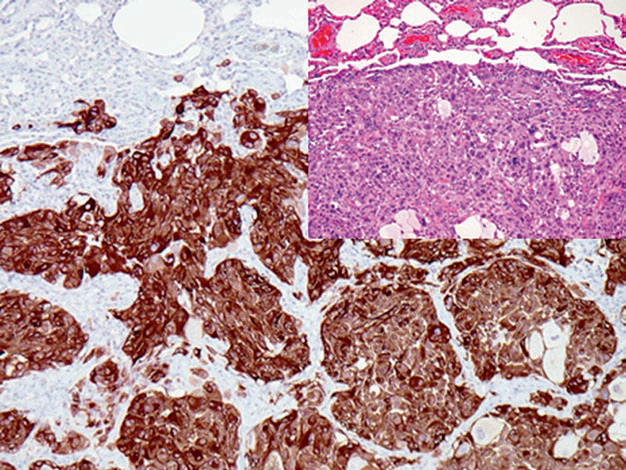
Figure 1. Metastatic malignant melanoma, HMB45 positive.
It should be noted that approximately 10% of malignant melanomas do not express HMB45, especially in amelanotic malignant melanoma, desmoplastic malignant melanoma, and spindle cell malignant melanoma, where this proportion is higher. The combined use of multiple malignant melanoma-related antibodies can significantly improve detection sensitivity, such as the combination of HMB45,Melan A, and tyrosinase. Additionally, some tumors with morphological similarities to malignant melanoma may also express HMB45, such as adrenal pheochromocytoma and clear cell tumors of the lung, but these tumors generally do not express tyrosinase or SOX-10.
Positive expression pattern: Cytoplasm
Recommended positive control tissue: Adrenal cortex
Melan AAlso known asMART-1, it is a melanocyte antigen and a member of the MAGE family involved in melanosome maturation and pigment expression in the endoplasmic reticulum of normal skin melanocytes and retinal cells. Tumors derived from these cells also express this antigen.
Melan Ais one of the most commonly used markers for malignant melanoma, expressed in over 90% of malignant melanomas. However, this marker is not specific for diagnosing malignant melanoma because it can also be expressed in other tumors, such as adrenal cortical tumors and sex cord-stromal tumors. The original author of this article suggests that it can be used as a screening antibody, and additional antibodies are needed to confirm the diagnosis of malignant melanoma.
Positive expression pattern: Cytoplasm
Recommended positive control tissue: Skin/Malignant melanoma
Tyrosinase catalyzes the synthesis of melanin from tyrosine in melanocytes. This is a very specific marker for malignant melanoma, but its positive rate in malignant melanoma is only over 80%, and its positivity is related to the tumor’s differentiation grade. Due to its high specificity, it is often used in combination with other malignant melanoma-related markers in immunohistochemical panels. Such panels are very effective in diagnosing epithelioid, desmoplastic, and spindle cell malignant melanoma and are helpful in detecting sentinel lymph node micrometastases.
Positive expression pattern: Nucleus
Recommended positive control tissue: Skin/Malignant melanoma
SOX-10 is a member of the SOX family of transcription factors and is a neural crest transcription factor involved in the maturation and differentiation of melanocytes and Schwann cells. Normally, SOX-10 is expressed in melanocytes, Schwann cells, and myoepithelial cells, and it is also a sensitive marker for various types of malignant melanoma, including the desmoplastic type.
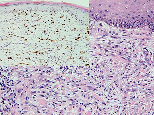
Figure 2. Desmoplastic malignant melanoma, immunohistochemistry for SOX-10 shows strong nuclear positivity.
Although SOX-10 is an excellent marker for malignant melanoma, it is not specific. Some other tumors may also show positive expression of this marker, such as schwannomas, neurofibromas, granular cell tumors, and up to 60% of malignant peripheral nerve sheath tumors. Additionally, SOX-10 is also a marker for triple-negative and metaplastic breast cancers. Myoepithelial cells and myoepithelial tumors also show strong positive expression of SOX-10, such as various types of salivary gland tumors. For suspicious cases, more specific malignant melanoma-related markers should be added to confirm or rule out the diagnosis.
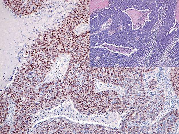
Figure 3. Triple-negative breast cancer, immunohistochemistry for SOX-10 shows moderate nuclear positivity in tumor cells.
WT-1 is another marker for malignant melanoma, which has been detailed in the mesothelioma section. Similar to HMB45, WT-1 helps differentiate malignant melanocytic lesions from benign melanocytic lesions.
MaiMai Recommendation: “Malignant melanoma is a highly malignant tumor, mostly occurring in the skin, but also seen at mucocutaneous junctions, ocular choroid, and leptomeninges. Its incidence is increasing year by year and has now become the leading fatal skin disease. Immunohistochemistry plays an important role in the definitive diagnosis of malignant melanoma when morphology is difficult to confirm! This issue of MaiMai recommends the following antibodies, you might want to try them.””
|
Antibody Name
|
Clone Number
|
Positive Control
|
Cellular Localization
|
|
Melan A
|
A103
|
Malignant melanoma
|
Cytoplasm
|
|
HMB45
|
HMB45
|
Malignant melanoma
|
Cytoplasm
|
|
SOX-10
|
EP268
|
Schwannoma, Malignant melanoma
|
Nucleus
|
|
WT-1*
|
MX012
|
Kidney tissue, Wilms tumor
|
Nucleus
|
*Marked as Maxin clone products
For more information, please contact: 800-8581156 or 400-889-9853
